про услугу
В каких случаях необходимо ультразвуковое исследование сердца, каковы его виды и как проводится? Разберемся в данной статье.
Что такое эхокардиография
Эхокардиография — это метод, который использует ультразвук для получения изображений сердца, сердечных клапанов и крупных сосудов.
Она является важным диагностическим тестом, поскольку она дает движущееся изображение сердца. С помощью ультразвука эхокардиография дает информацию о форме, размере, функции, ритме сердца, движении и толщине его стенок, а также о состоянии околосердечной сумки и функционировании клапанов.
Кроме того, он может предоставить информацию о малом круге кровообращения и его давлении, начальном отделе аорты и увидеть, есть ли жидкость вокруг сердца (перикардиальный выпот).
Эхокардиография полезна для получения информации о функционировании сердца как в состоянии покоя, так и после физической нагрузки или приема лекарственных препаратов ( стресс ЭхоКГ).
Благодаря использованию ультразвука эхокардиография представляет собой тот вид исследования, который не облучает пациента (в отличие от рентгена, КТ и т. д.). Поэтому для данного вида диагностики не существует абсолютных противопоказаний.
Изображения эхокардиографии можно получить в разных режимах:
-
М или одномерный режим: в этом случае показывается узкая часть сердца.
-
Двумерное или 2D: предлагает изображение анатомии сердца (позволяет видеть различные структуры) во время движения.
-
Цветной допплер: позволяет увидеть кровоток в сердце и артериях и измерить его.
-
3D: полученные изображения имеют трехмерное изображение. Оно создается из нескольких двухмерных изображений.
Есть и другие информативные режимы эхокардиографии: импульсный допплер, непрерывный допплер и т. д. Наиболее часто используемый метод — двумерное эхо.
Показания для проведения УЗИ сердца
Выделим наиболее частые показания для ультразвукового исследования сердца:
-
Стенокардия.
-
Сердечные аритмии.
-
Врожденный порок сердца.
-
Клапанные заболевания.
-
Острый инфаркт миокарда.
-
Сердечная недостаточность.
-
Артериальная гипертензия.
-
Заболевания перикарда.
-
Эндокардит.
-
Подозрение на тромбоэмболию.
-
Подозрение на опухолевое образование.
Диагностическое применение эхокардиографии
Остановимся подробнее на основных вариантах диагностического применения УЗИ сердца.
-
Ишемическая болезнь сердца
Это патологическое состояние, характеризующееся поражением сердечной мышцы вследствие нарушенного кровотока (ишемии). Транспорт крови к сердцу происходит по коронарным артериям, и если их просвет сужается вследствие атеросклероза, развивается ишемия с появлением характерных симптомов.
В этих случаях полезна эхокардиография, поскольку она позволяет выявить измененную функцию мышцы во время цикла сокращения и расслабления.
-
Клапанные патологии
Пороки клапанов сердца – это заболевания, обусловленные структурными аномалиями или нарушениями функции клапанов сердца.
Клапаны – это анатомические структуры, служащие для обеспечения движения крови по камерам сердца только в одном направлении.
Во время сердечного цикла клапаны играют фундаментальную роль в координации и обеспечении однонаправленности кровотока, проходящего через сердце.
В случае диагностики функции желудочков УЗИ раскрывает свой максимальный аналитический потенциал.
-
Кардиомиопатии
Кардиомиопатия – общий термин, используемый для обозначения патологий, которые в первую очередь поражают сердечную мышцу, вызывая снижение ее способности сокращаться и расслабляться.
И в этом случае эхокардиография играет основополагающую и незаменимую роль как в диагностике, так и в ведении больного в динамике.
Кардиомиопатии разделяют на три основных типа:
-
дилятационная (наиболее распространенная): в этом случае левый желудочек расширяется и не может перекачивать кровь по всему телу так же эффективно, как здоровое сердце;
-
гипертрофическая: сердце имеет утолщенные стенки и маленькие камеры сердца;
-
рестриктивная (самый редкий тип): стенки камер сердца становятся жесткими, что нарушает расслабление после сокращения, и это не позволяет сердцу завершить свой цикл.
-
-
Патологии аорты
Аневризмы грудной аорты представляют собой аномальное расширение аорты над диафрагмой.
Эхокардиография имеет решающее значение в диагностике расслаивающей аневризмы и используется для установления ее точных размеров. Также она незаменима в диагностике врожденных пороков сердца, выявлении образований сердца и патологии перикарда.
Виды эхокардиографии
Существуют различные варианты проведения ультразвукового исследования. Рассмотрим характеристики каждого из них.
-
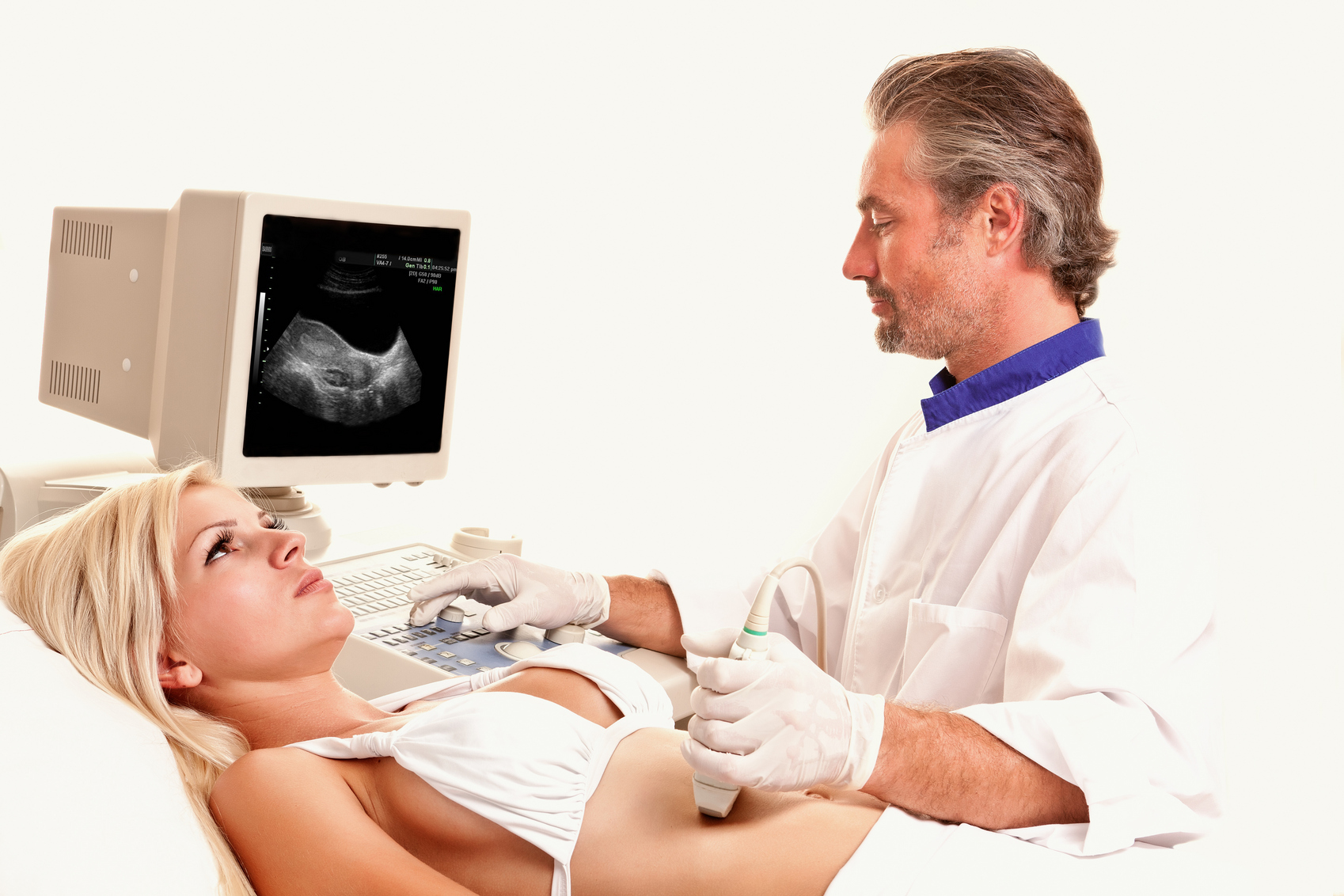
Трансторакальная эхокардиография
Этот вид используется чаще всего. Для визуализации сердца датчик помещают на грудь пациента, обычно с левой стороны.
-
Чреспищеводная эхокардиография
Она заключается в визуализации сердца с помощью датчика, соединенного с зондом или трубкой, который вводится через рот в пищевод, откуда можно получить изображения сердца. Следовательно, это инвазивный метод.
Обычно это дополнительное исследование к трансторакальной ЭхоКГ, которая дает аналогичную, но более подробную информацию об определенных структурах, таких как сердечные клапаны. Кроме того, это помогает исключить наличие тромбов (сгустков), опухолей или врожденных пороков сердца, которые невозможно полностью обнаружить с помощью трансторакальной эхокардиографии.
За 4-6 часов до чреспищеводного УЗИ нельзя употреблять пищу и жидкости. Также не следует принимать какие-либо пероральные лекарства в этот период. Предпочтительно, чтобы пациента сопровождал член семьи или друг, поскольку иногда для проведения теста необходимо ввести успокоительное. Если у пациента есть съемные зубные протезы,их необходимо снять во время обследования.
Продолжительность исследования - 45-60 минут.
После не следует есть какую-либо пищу в течение примерно 2 часов. Также не желательно садиться за руль в течение 2-4 часов после исследования, если седативное средство было введено внутривенно. Пожилых людей необходимо сопровождать в течение следующих 2-4 часов.
-
Стресс-эхокардиография
Она заключается в визуализации сердца с помощью ЭхоКГ в условиях физической или лекарственной нагрузки.
Твердую пищу нельзя употреблять за 2 часа до исследования. Врач, который рекомендует данное исследование, сообщит пациенту, следует ли ему принимать обычное лекарство или необходимо приостановить его прием.
После стресс-ЭхоКГ можно сразу же вернуться к повседневным делам.
-
Фармакологическая стресс-эхокардиография
Она заключается в визуализации сердца с помощью ультразвука при введении препарата, который заставляет сердце работать быстрее и интенсивнее. Это неинвазивный тест, сочетающий использование трансторакальной эхокардиографии с введением лекарственного препарата. Обычно используется добутамин. Он увеличивает частоту сердечных сокращений и силу сокращения сердца (среди других эффектов).
Так, с помощью данного теста можно стимулировать сердце и увидеть, как оно работает во время усилий (когда мы двигаемся или тренируемся). Изображения сначала будут получены при состоянии покоя сердца, а затем после введения препарата.
Цель этого исследования — выяснить, есть ли изменения в сокращении стенок сердца, которые в большинстве случаев являются вторичными по отношению к ишемической болезни сердца.
Среди тех, кому показана фармакологическая ЭхоКГ:
-
Пациенты, у которых был проведен стресс-тест с подозрением на ишемическую болезнь сердца, и результат оказался неубедительным.
-
Пациенты, у которых уже был сердечный приступ и у которых желательно оценить риск.
-
Пациенты с уже известными поражениями коронарных артерий и перед их лечением необходимо выяснить, как эти поражения влияют на функцию сердца.
Пациенту не следует употреблять твердую пищу за несколько часов до исследования. Как правило, если пациент принимает бета-блокаторы, необходимо приостановить их прием за 1-3 дня до проведения исследования. Но в любой ситуации важно следовать рекомендациям лечащего врача.
Если доктор не посоветует иного, сразу после обследования можно вернуться к работе или домашним делам.
-
Контрастная эхокардиография
В этом случае используется обычная эхокардиография, но прежде через вену вводится специальный контраст, позволяющий значительно лучше рассмотреть структуры сердца и сосудов.
Никакой специальной подготовки это не требует.
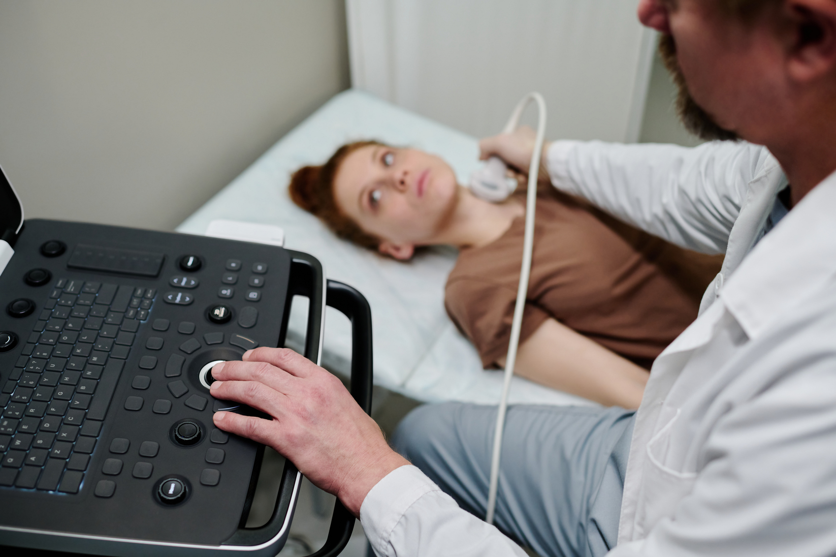
Как проводится УЗИ сердца
Если речь идет о классической, трансторакальной ЭхоКГ, проводящий гель наносится либо на грудь пациента, либо непосредственно на датчик. Кардиолог будет перемещать датчик по груди пациента, чтобы получить различные изображения.
Пациент в процессе лежит и остается максимально спокойным. Эхокардиограмма безболезненна и не вызывает каких-либо побочных эффектов.
Исследование обычно длится от 15 до 30 минут.
-
Чреспищеводная эхокардиография
Перед ее проведением медсестра может дать пациенту пастилку для рассасывания или обезболивающий спрей для горла. Когда пациент ляжет на кушетку, обнажив грудь, на ней разместят наклейки с электродами для просмотра электрокардиограммы во время исследования. Медсестра также может ввести в вену седативное лекарство.
Затем кардиолог поместит трубку в рот пациента и попросит его проглотить, прежде чем вводить ее в пищевод, что может вызвать некоторую тошноту, которая пройдет, как только трубка окажется внутри. В дальнейшем будет проведено исследование, во время которого, если было введено успокоительное средство, пациент настолько расслабится, что может даже заснуть. После возможно легкое головокружение.
Анестетик для горла может вызвать кашель. К тому же, при извлечении зонда рекомендуется кашлять, чтобы удалить скопившуюся мокроту.
-
Стрессовая ЭхоКГ
На грудь пациента будут помещены электроды для просмотра электрокардиограммы и манжета на руку для измерения артериального давления. Далее следует подняться на беговую дорожку или велотренажер, где пациент будет ходить несколько минут. После нужно будет вернуться в лежачее положение. Кардиолог будет делать записи на эхокардиографе перед выполнением упражнения, во время его выполнения и сразу после его окончания.
Во время исследования могут появиться боли в груди, усталость или дискомфорт, которые исчезают после. О подобных ощущениях необходимо сообщить своему врачу.
Примерная продолжительность исследования составляет 30-60 минут.
-
Фармакологическое стресс-УЗИ
В этом случае пациенту поставят капельницу. Затем на грудь будут помещены электроды для просмотра электрокардиограммы во время исследования. Кроме того, оденут манжеты для измерения артериального давления.
Тест состоит из четырех фаз: когда сердце находится в состоянии покоя, при низких и высоких дозах лекарственного препарата и фазы восстановления. Как только будут получены нормальные изображения эхокардиографии (в покое, без препарата), во время введения препарата будет сделано несколько эхокардиографических записей.
Возможны покалывание лица, жар в лице, головная боль, тремор, головокружение, тошнота, учащенное сердцебиение (которые почти всегда присутствуют из-за увеличения частоты сердечных сокращений), усталость или боль в груди в течение нескольких секунд. О любом дискомфорте или симптомах следует сообщать врачу. Ориентировочная продолжительность исследования - 45 минут.
-
Контрастная эхокардиография
Пациенту установят электроды для просмотра ЭКГ во время исследования. Затем, пока медсестра делает несколько инъекций контраста в вену, датчик будет помещен в разные части грудной клетки, чтобы исследовать сердце и клапаны с разных плоскостей. Укол обычно незаметен, поэтому не стесняйтесь сообщать о любых странных ощущениях. Во время исследования может быть слышен некоторый шум, соответствующий скорости движения крови внутри сердца. После завершения исследования медсестра наложит повязку или лейкопластырь на область введения контраста.
Примерная продолжительность исследования - 30-60 минут.
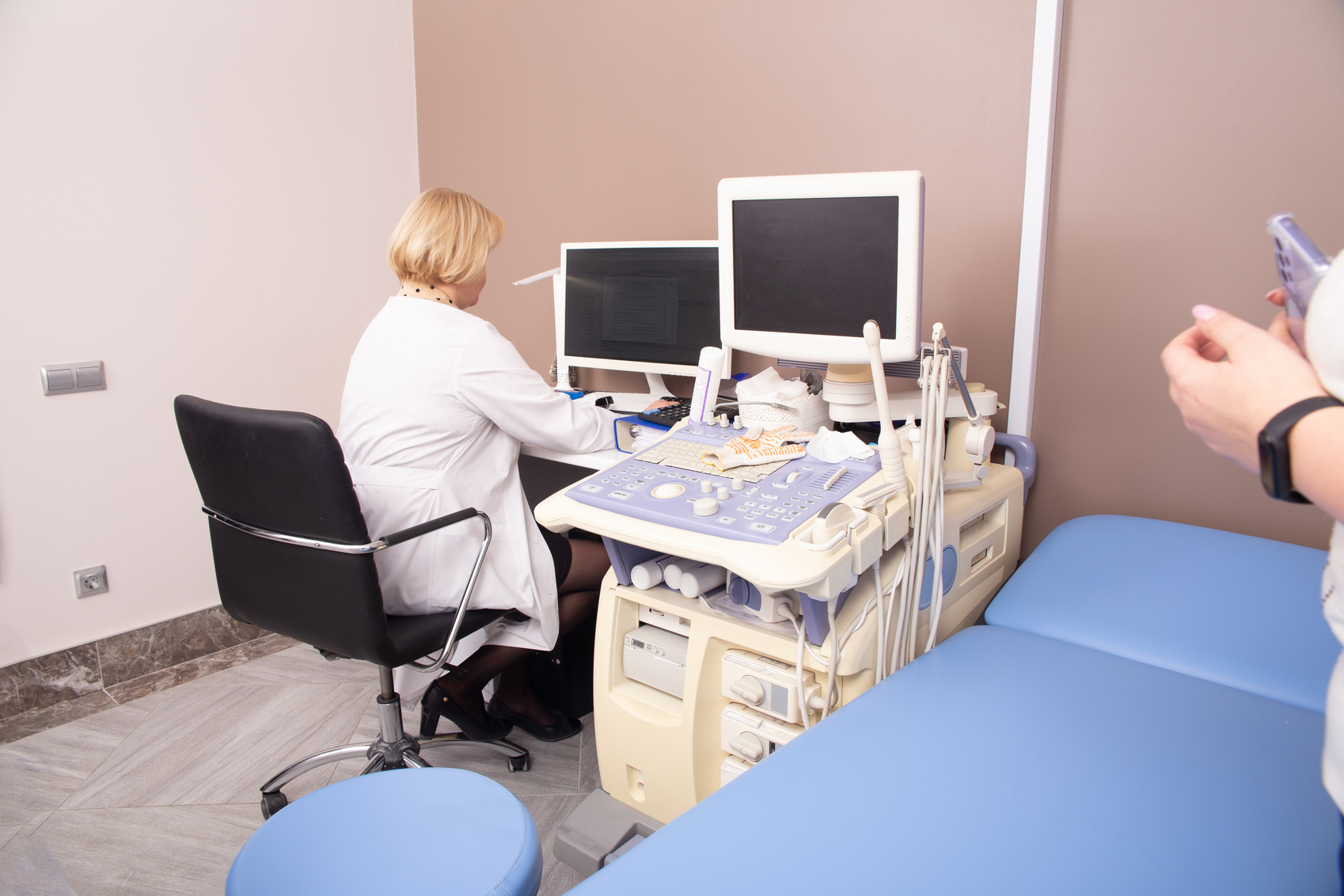
Преимущества УЗИ сердца в клинике Expert Clinics
В Expert Clinics применяется новейшее диагностическое оборудование, которое дает точные результаты. Также в клинике работают врачи, обладающие знаниями из области антивозрастной медицины.
Поставив диагноз, они будут тщательно работать не только над устранением симптомов, но также над поиском глубинной причины “сбоя” в организме. Способы коррекции различных патологий подбираются строго персонализировано, но всегда максимально эффективно.
Краткие выводы
-
УЗИ сердца - важный диагностический инструмент для определения состояния сердца и его функций.
-
Эхокардиография использует ультразвуковые волны для получения изображений сердечной мышцы, клапанов и сосудов.
-
УЗИ сердца полезно для оценки функционирования сердца в состоянии покоя и после физических нагрузок.
-
Отсутствие противопоказаний для проведения УЗИ делает его безопасным и широко используемым методом диагностики.
-
Различные режимы УЗИ сердца позволяют получить информацию обо всех аспектах работы сердца.
-
Показания для УЗИ сердца включают стенокардию, аритмии, врожденные пороки сердца, клапанные заболевания, инфаркт миокарда, сердечную недостаточность и артериальную гипертензию и так далее.
-
В клинике Expert Clinics используются новейшие технологии и работают врачи с опытом в антивозрастной медицине, что позволяет им проводить точную индивидуальную диагностику и назначать оптимальное лечение, которое дает быстрые результаты.
Список использованной литературы
- Arntfield R, Pace J, Hewak M, Thompson D. Focused Transesophageal Echocardiography by Emergency Physicians is Feasible and Clinically Influential: Observational Results from a Novel Ultrasound Program. J Emerg Med. 2016 Feb;50(2).
-
Nazerian P, Vanni S, Castelli M, Morello F, Tozzetti C, Zagli G, Giannazzo G, Vergara R, Grifoni S. Diagnostic performance of emergency transthoracic focus cardiac ultrasound in suspected acute type A aortic dissection. Intern Emerg Med. 2014 Sep;9(6):665-70.
-
Lang RM, Badano LP, Mor-Avi V, Afilalo J, Armstrong A, Ernande L, Flachskampf FA, Foster E, Goldstein SA, Kuznetsova T, Lancellotti P, Muraru D, Picard MH, Rietzschel ER, Rudski L, Spencer KT, Tsang W, Voigt JU. Recommendations for cardiac chamber quantification by echocardiography in adults: an update from the American Society of Echocardiography and the European Association of Cardiovascular Imaging. Eur Heart J Cardiovasc Imaging. 2015 Mar;16(3):233-70.
-
Kennedy Hall M, Coffey EC, Herbst M, Liu R, Pare JR, Andrew Taylor R, Thomas S, Moore CL. The "5Es" of emergency physician-performed focused cardiac ultrasound: a protocol for rapid identification of effusion, ejection, equality, exit, and entrance. Acad Emerg Med. 2015 May;22(5):583-93.
-
Blaivas M. Incidence of pericardial effusion in patients presenting to the emergency department with unexplained dyspnea. Acad Emerg Med. 2001 Dec;8(12):1143-6.
-
Secko MA, Lazar JM, Salciccioli LA, Stone MB. Can junior emergency physicians use E-point septal separation to accurately estimate left ventricular function in acutely dyspneic patients? Acad Emerg Med. 2011 Nov;18(11):1223-6.
-
Iung B, Rouzet F, Brochet E, Duval X. Cardiac Imaging of Infective Endocarditis, Echo and Beyond. Curr Infect Dis Rep. 2017 Feb;19(2):8.
СВЯЗАННЫЕ КОМПЛЕКСНЫЕ ПРОГРАММЫ