про услугу
УЗИ органов мошонки у мужчин — информативный метод диагностики, позволяющий визуализировать органы мошонки и выявлять наличие патологий. Для исследования используются высокочастотные ультразвуковые волны.
В переводе с латинского мошонка означает «мешочек». Внутри нее находятся семенники — парные органы, в которых происходит образование сперматозоидов и тестостерона. Это важная часть репродуктивной системы, для защиты которой природа предусмотрела специальное кожно-мышечное образование.
Мужчины в большинстве случаев игнорируют отеки и другие изменения половых органов, даже боль. Это приводит к развитию патологий и осложнений, когда лечение становится трудным или невозможным. Поэтому важно при наличии проблем в этой области, а также периодически с целью профилактики проходить обследование.
Ультразвуковое исследование мошонки наряду с пальпацией и визуальным осмотром является методом первичной диагностики. Он позволяет визуализировать контуры, размеры, расположение и строение внутренних органов, состояние тканей, сосудов. Это помогает установить или исключить наличие патологий.
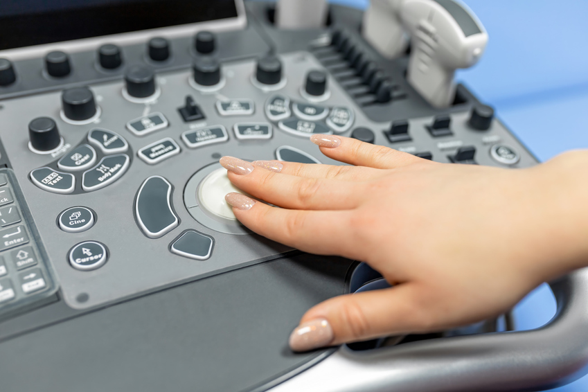
Используются компактные приборы, излучающие ультразвуковые волны частотой 5–10 мегагерц. Глубины сканирования 10 см достаточно для исследования органов, а их размеры определяются с точностью до 1 мм (фиксируются даже незначительные отклонения в размерах).
Также применяется отдельная методика УЗИ сосудов мошонки — ультразвуковая допплерография. Она позволяет визуализировать все кровеносные сосуды, направление и скорость движения крови. Это отличный способ диагностики сосудистых заболеваний, позволяющий точно установить их причину. Можно детально рассмотреть сужение сосудов, бляшки и тромбы. Методика используется при варикозном расширении вен.
Преимущества ультразвуковой диагностики
Безопасный, быстрый и высокоточный метод диагностики широко применяется в клиниках всего мира. Он позволяет контролировать состояние мужского здоровья, своевременно выявлять патологии. Мужчинам рекомендуется проходить обследование ежегодно в целях профилактики.
Перечислим главные достоинства метода:
-
Безболезненность, неинвазивность. Хирургического вмешательства не требуется.
-
Безопасность. Ультразвуковые волны в используемом диапазоне не вредят органам и клеткам.
-
Не требуется стимуляции эрекции, инъекций контрастных препаратов.
-
Быстрота. Результаты можно получить уже через 20 минут.
-
Картинка в реальном времени идеально подходит для проведения операции, биопсии.
-
Отсутствие возрастных ограничений и противопоказаний. Процедура назначается даже маленьким детям и пожилым людям.
-
Широкая доступность, низкая стоимость.
УЗИ имеет только один недостаток — невозможно отличить доброкачественное образование от злокачественного. Для этого необходимы дополнительные исследования.
Показания к проведению УЗИ
Любые изменения во внешнем виде органа и появление неприятных ощущений — повод сделать УЗИ мошонки.
На необходимость обследования могут указывать следующие признаки:
-
боль в яичках из-за травмы или воспаления;
-
появление твердых образований — могут говорить о наличии опухоли, кисты;
-
механические повреждения мошонки: ушибы, царапины, порезы;
-
объемное образование, которое прощупывает сам пациент;
-
варикозное расширение вен;
-
увеличение/ уменьшение яичек;
-
монорхизм (врожденное отсутствие одного яичка);
-
крипторхизм (расположение яичек вне мошонки), они могут находиться в паховом канале или брюшной полости);
-
замедленное или преждевременное половое созревание;
-
изменения в спермограмме;
-
появление крови в сперме;
-
проявления эректильной дисфункции.
Какие заболевания диагностируются при исследовании
УЗИ органов мошонки проводится мужчинам с целью выявления патологий семенников, придатков и сосудов.
Этот вид обследования хорошо визуализирует следующие патологические процессы:
-
Эпидидимит (воспаление придатков яичек).
-
Перекрут яичка. Опасное состояние, при котором происходит скручивание семенного канатика, в связи с чем кровоснабжение яичка прекращается. Наиболее часто встречается у подростков в возрасте от 10 до 16 лет.
-
Кисты. Являются одним из самых распространенных заболеваний мошонки.
-
Опухоли. По данным журнала «Онкоурология», ежегодно в мире регистрируют около 75 тыс. случаев заболеваний раком яичек. В зоне риска — мужчины от 45 до 50 лет.
-
Гидроцеле (водянка). Это патология, при которой вокруг яичек накапливается лишняя прозрачная жидкость.
-
Гематоцеле. Скопление крови в полости между оболочками яичке. Причиной могут стать неудачная пункция, травма (разрыв, перекрут), повышенная кровоточивость, спонтанные кровотечения.
-
Лимфедема (избыточное скопление богатой белком жидкости в тканях).
УЗИ позволяет определить структуру, форму, размер и работу органов мошонки, а также функционирование кровеносных сосудов.
Подготовка к УЗИ
Если для некоторых других видов УЗИ требуется соблюдение диеты и наполнение мочевого пузыря, то здесь не требуется предварительной подготовки.
-
Достаточно простой гигиены половых органов. По желанию можно сбрить волосы в области паха — так проще будет убрать остатки геля.
-
За 1–2 часа до посещения клиники рекомендуется воздержаться от курения. Никотин сужает сосуды, что влияет на результаты исследования.
-
Направляясь на обследование, не забудьте взять с собой результаты предыдущих анализов и обследований.
Если процедуру предстоит пройти мальчику, родителям стоит морально его подготовить. Если ребенок будет напуган или взволнован, яички втянутся в паховые каналы, что затруднит диагностику.
Как проходит УЗИ паховой области
Исследование занимает в среднем 15–20 минут. Если у специалиста появятся подозрения на патологические процессы, то процедура может продлиться до получаса. Диагностика является абсолютно безболезненной.
-
Мужчина заходит к врачу, раздевается, максимально подробно рассказывает о своих жалобах.
-
Далее пациент берет одноразовую пеленку, застилает ей кушетку и ложится на спину.
-
Лежа на спине, необходимо в одном положении зафиксировать половой член, и, не надавливая, опустить его на живот, придерживая рукой.
-
Доктор смазывает датчик гелем комнатной температуры и начинает процедуру.
-
Врач делает оценку параметров яичек и придатков (структуру, размер, толщину стенок), фиксирует наличие отклонений.
После процедуры пациенту выдается заключение с подробным комментарием и пожеланиями к дальнейшим действиям.
Расшифровка результатов
По окончании процедуры пациент получает протокол с заключением, где фиксируются результаты обследования мошонки в трех проекциях: продольной, поперечной и косой.
Эти результаты нужно показать лечащему врачу для расшифровки, постановки диагноза и последующего назначения лечения.
В нашей клинике вы можете не только пройти диагностику, но и записаться на прием к профильному специалисту.
Какие показатели УЗИ мошонки считаются нормальными
В протоколе ультразвукового исследования содержатся следующие пункты:
-
Параметры (форма, размеры) яичек и их придатков. Нормальные размеры яичек: длина — до 5 см, ширина — до 3 см в ширину, толщина — до 2 см.
-
Скопление жидкости между стенками мошонки и яичка (в норме 1–2 мл);
-
Толщина стенки (до 8 мм).
В норме яички имеют овальную форму, и размеры могут отличаться. Обычно правое находится немного выше левого. Контуры органов должны быть четкими. Эхоплотность (способность отражать ультразвук) — средняя.
Преимущества УЗИ в Expert Clinics
В медицинском центре Expert Clinics вы можете пройти ультразвуковую диагностику мужских половых органов.
Обследование и лечение в нашей клинике предполагает множество преимуществ:
-
Врачи Expert Clinics — специалисты международного уровня, постоянно совершенствующие свои навыки в России и за рубежом. Они обладают знаниями из области превентивной антивозрастной медицины, что позволяет им выявлять и эффективно устранять истинные причины различных сбоев в работе организма.
-
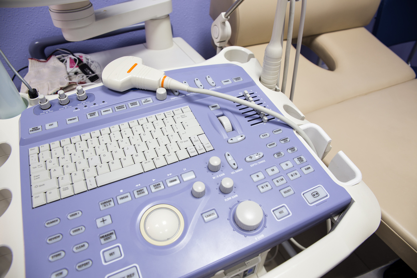
Самое качественное и современное оборудование. Высокоточные аппараты для ультразвуковой диагностики в сочетании с высочайшим уровнем знаний врачей позволяют проводить обследования с максимальной достоверностью.
-
Комфортные условия и исключительно индивидуальный подход к каждому пациенту.
-
Комплексный подход, предусматривающий работу с пациентом на всех этапах: от консультации врача и сдачи анализов до полного лечения вашего заболевания.
-
Разумные и прозрачные цены на медицинские услуги.
-
Удобное расположение клиники в центре Москвы.
Записаться на УЗИ мошонки можно по телефону или на сайте. Оставьте свои контакты, и оператор свяжется с вами для уточнения деталей.