про услугу
В каких случаях необходимо пройти данное исследование и как оно проводится?
Что такое УЗИ вен ног?
Ультразвуковое исследование вен нижних конечностей (допплерография) - это фундаментальное исследование сосудов для оценки кровообращения, поскольку ноги отвечают за отток жидкостей из нижней части тела к сердцу.
В этом исследовании используются два типа различных технологий: ультразвуковое исследование и эффект Доплера.
-
Ультразвук — это метод визуализации, использующий физическое явление излучения и приема ультразвукового луча, который, воздействуя на ткани, создает различное сопротивление прохождению волн, обеспечивая получение изображений в зависимости от плотности самой ткани.
-
Эффект Доплера — это физический принцип, когда ультразвуковые волны во время прохождения сталкиваются с движущейся структурой, которой в случае исследования сосудов является кровь.
Таким образом, с помощью наружного датчика и геля в качестве проводника, благодаря этим двум методам можно получить морфологическую информацию о структуре, стенке и ходе кровеносного сосуда, а также функциональную информацию о скорости кровотока и направления крови.
Так, доктора могут выявлять любые закупорки, стенозы (сужения), атеросклеротические бляшки или другие аномалии, которые могут повлиять на кровообращение.
Какие патологии выявляет УЗИ вен нижних конечностей
Мониторинг состояния кровеносных сосудов с помощью ультразвуковой допплерографии имеет важное значение для выявления потенциально серьезных патологий, поражающих сосудистую систему, в частности вен и артерий.
Эти два типа кровеносных сосудов выполняют разные функции и имеют разные структурные характеристики, поэтому различают артериальную и венозную допплерографию:
Артериальная допплерография обычно используется для проверки состояния здоровья крупнейших кровеносных сосудов (точнее, артерий), которые содержат кровь, насыщенную кислородом и питательными веществами, которая, начиная с левого желудочка сердца, достигает всех других органов.
Однако венозная допплерография фокусируется на наблюдении кровеносных сосудов, которые следуют обратным путем, то есть, транспортируют кровь, насыщенную углекислым газом, из периферических участков в правое предсердие.
С помощью этого диагностического исследования можно выявить следующие патологии:
-
Аневризмы венозных стенок. Они представляют собой аномальные и постоянные расширения стенки вены, которые могут быть вызваны травмой или морфологическими изменениями. Когда расширение достигает критического уровня, сосуд может разорваться, что приведет к внутреннему кровотечению.
-
Стеноз или окклюзия. Это аномальное и неестественное сужение кровеносного сосуда, способное препятствовать нормальному току крови, тогда как венозная окклюзия заключается в остановке кровотока внутри вены.
-
Венозная недостаточность. Это патологическое состояние кровообращения, при котором вены больше не могут транспортировать нужное количество крови от конечностей к сердцу.
-
Варикозное расширение вен. Это заболевание, характеризующееся постоянным расширением вены, которая кажется извилистой и видима невооруженным глазом. Изменения связаны с несостоятельностью клапанов и нарушением оттока крови из вен нижних конечностей.
-
Тромбоз. Это сосудистая патология, возникающая в результате формирования тромба внутри вены. Тромб – это плотная масса, образующаяся в результате свертывания крови в сердечно-сосудистой системе.
-
Флебит - воспаление вены, которое в большинстве случаев сопровождается наличием тромба.
Поскольку, как уже упоминалось, кровеносные сосуды нижних конечностей имеют основополагающее значение для оттока крови из нижней части тела к сердцу, ультразвуковая допплерография является важным диагностическим исследованием, позволяющим оценить состояние этих сосудов и выявить любые потенциально опасные расстройства.
Показания к УЗИ вен нижних конечностей
Ультразвуковое исследование нижних конечностей рекомендуется в следующих ситуациях:
-
Болевые ощущения и ощущения усталости. Если вы испытываете постоянную боль, онемение мышц, тяжесть или судороги в ногах, вам может потребоваться обследование для оценки состояния артерий и вен.
-
Изменение цвета кожи над сосудами или их внешнего вида (выпячивания, узелки), появление сосудистых звездочек.
-
Тромбоз, тромбофлебит, атеросклероз. Люди с проблемами кровообращения, такими как вышеперечисленные или синдром беспокойных ног или хроническая венозная недостаточность, могут воспользоваться этим инструментом для оценки тяжести своего состояния. Венозная недостаточность обычно проявляется в виде варикозного расширения вен. При поражении системы глубоких вен обычными клиническими проявлениями являются: ощущение тяжести в ногах, боль, отек (отечность) лодыжек и голеней, жжение. Все это обычно происходит днем или ночью и менее заметно утром после пробуждения.
-
Необъяснимый отек. Если ваши ноги отекают по необъяснимой причине, вам может потребоваться УЗИ сосудов нижних конечностей.
-
Язвы или раны на ногах. Если на ногах имеются стойкие язвы или раны, которые плохо заживают, этот метод может помочь определить, нарушено ли кровообращение в данной области.
-
Послеоперационный осмотр. После сосудистой хирургии часто используется цветное допплеровское картирование, чтобы контролировать успех операции и ход заживления.
-
Профилактика. В некоторых случаях лечащий врач может порекомендовать УЗИ в рамках профилактического обследования. Это особенно актуально для пациентов с сосудистыми факторами риска, такими как диабет, курение, гипертония или гиперхолестеринемия.
ВАЖНО: в случаях подозрения на тромбоз глубоких или поверхностных вен важно диагностировать пораженные вены и степень проблемы. Тромбы в венах ног часто являются причиной опасных для жизни тромбоэмболий легочной артерии. Первоначальная диагностика при своевременном лечении снижает риск развития осложнений.
Как проходит исследование
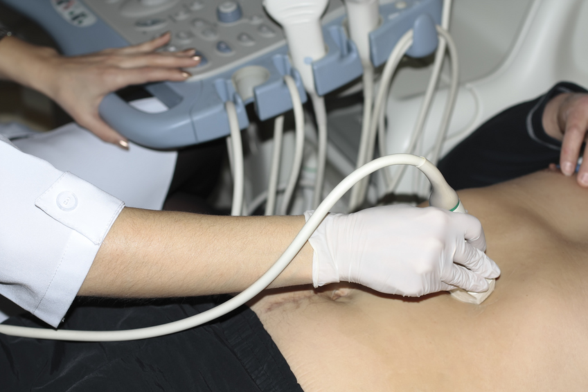
Специалист нанесет гель на водной основе на обследуемый участок ног. Он улучшает контакт между ультразвуковым датчиком и кожей, обеспечивая лучшую передачу звуковых волн.
Полное исследование поверхностной и глубокой венозной системы голени для выявления и характеристики венозной недостаточности заключается в осмотре больного лежа и стоя - доктор будет перемещать датчик в разных направлениях. Это обеспечивает полную визуализацию сосудов ног.
Звуковые волны, посылаемые зондом, проходят через ткани тела и отражаются от кровеносных сосудов. Зонд обнаруживает отраженные волны и преобразует их в изображения в реальном времени на экране.
Обследование начинается от паха и заканчивается на лодыжке и обычно занимает 30-60 минут.
Во время обследования специалист оценит диаметр кровеносных сосудов, скорость кровотока и наличие каких-либо препятствий или аномалий. Эта информация необходима для точной диагностики.
Подготовка пациента
Никакой специальной подготовки к проведению УЗИ вен нижних конечностей не требуется. Доктор может рекомендовать отказаться от употребления кофеина и венотоников непосредственно перед исследование.
Также перед допплерографией следует снять украшения и другие металлические предметы, которые могут мешать ультразвуку.
Безопасно ли УЗИ ног?
Данная методика считается безопасной и неинвазивной. Она не требует ионизирующего излучения, как рентгеновские лучи, и хорошо переносится большинством пациентов. Поэтому абсолютных противопоказаний не существует.
Тем не менее, важно сообщать специалисту о любых аллергиях или чувствительности к давлению или гелю, используемому во время исследования.
Преимущества УЗИ вен нижних конечностей в Expert Clinics
Если вы хотите пройти допплерографию нижних конечностей, вы можете положиться на профессионализм специалистов клиники Expert Clinics, которая предлагает высококачественные услуги диагностической визуализации, выполняемые квалифицированным персоналом с использованием новейшего оборудования.
В клинике установлены ультразвуковые системы последнего поколения, которые гарантируют высокую точность и отличное разрешение изображений.
Интерпретировать результаты могут наши доктора, обладающие знаниями из области антивозрастной медицины. Они способны заметить патологию в самом начале ее развития и вовремя купировать проблему.