
про услугу
Кондиломы — это образования, вызванные вирусом папилломы человека (ВПЧ), которые нередко возникают на слизистых оболочках половых органов, анальной области и других интимных зонах. Эти новообразования могут причинять физический дискомфорт и вызывать психологический стресс.
Современная медицина предлагает различные способы удаления кондилом, и коагуляция — один из наиболее эффективных и щадящих методов. В этой статье мы рассмотрим, что представляет собой данная процедура, каковы ее разновидности, как проходит подготовка и реабилитация, а также разберём её преимущества перед другими методами лечения.
Что такое кондиломы и почему их нужно удалять?
Кондиломы — это доброкачественные разрастания тканей, вызванные вирусом папилломы человека (ВПЧ). Они чаще всего возникают на слизистых оболочках наружных и внутренних половых органов — в области половых губ, влагалища, шейки матки, а также в анальном канале и на коже вокруг заднего прохода. По внешнему виду кондиломы напоминают наросты или бородавки телесного или розоватого цвета. Эти образования могут быть как единичными, так и множественными, нередко сливаются между собой и формируют гроздевидные структуры.
Кондиломы доставляют не только физический, но и психологический дискомфорт. Они могут вызывать зуд, жжение, болезненность при половом акте, раздражение кожи и слизистых оболочек, а также кровоточивость при травматизации. Особенно опасно механическое повреждение кондилом в интимной зоне: оно может спровоцировать воспаление, вторичную бактериальную инфекцию и даже привести к развитию дисплазии слизистой оболочки, что повышает риск онкологических заболеваний в будущем.
Удаление кондилом — важная часть комплексного лечения ВПЧ-инфекции. Это помогает снизить вероятность их дальнейшего распространения, уменьшить риск осложнений и улучшить качество жизни пациента. Кроме того, удаление снижает риск передачи вируса половым партнерам, что особенно актуально для планирующих беременность.
Методы удаления кондилом
Для удаления кондилом в клинической практике применяются различные современные методы, выбор которых зависит от размеров, количества, локализации образований и общего состояния здоровья пациента. Основные методы:
- Химическая деструкция — воздействие на кондиломы специальными составами (кислотами, щелочами, фенолами), которые вызывают разрушение тканей.
- Криодеструкция — замораживание патологических образований с помощью жидкого азота, что приводит к их некрозу и последующему отторжению.
- Электрокоагуляция — использование высокочастотного тока для прижигания и удаления кондилом.
- Радиоволновая энуклеация — бесконтактное удаление с помощью радиоволн (аппараты «Сургитрон», «Фотек»).
- Лазерная коагуляция — воздействие лазером (CO₂-лазер или диодный лазер) для точечного удаления кондилом с минимальной травматизацией тканей.
Каждый метод имеет свои показания и ограничения, поэтому окончательный выбор всегда делает врач после осмотра пациента. При принятии решения учитываются такие факторы, как площадь поражения, склонность к рецидивам, сопутствующие заболевания, наличие беременности и даже психологический настрой пациента.
Что такое химическая коагуляция
Химическая коагуляция — это метод точечного разрушения патологических тканей с помощью специально подобранных химических соединений. Препараты, применяемые для процедуры, содержат высокоактивные кислоты (например, трихлоруксусную, азотную), щёлочи или органические соединения (например, фенол), способные вызывать прижигающий эффект. Под их действием белки тканей сворачиваются (коагулируются), что приводит к гибели и последующему отторжению кондиломы.
Механизм воздействия основан на контролируемом химическом ожоге ограниченной глубины, после которого начинается естественный процесс заживления. Правильно выполненная процедура затрагивает исключительно измененные ткани, не повреждая здоровые участки.
Метод применяется, как правило, при единичных или небольших по размеру кондиломах, особенно в случаях, когда другие способы временно недоступны или противопоказаны. Он может быть использован как самостоятельный способ лечения или как дополнительный этап после хирургического удаления.
Ключевые особенности метода:
- процедура проводится без применения аппаратов и занимает всего несколько минут;
- не требует анестезии в большинстве случаев;
- подходит для амбулаторного применения под контролем врача;
- важно строго соблюдать технику нанесения препарата, чтобы избежать ожога окружающей ткани.
Химическая коагуляция широко применяется в гинекологии и дерматологии благодаря своей простоте и доступности, однако требует осторожного подхода, особенно при обработке слизистых оболочек. Эффективность метода во многом зависит от опыта специалиста и правильного подбора состава.
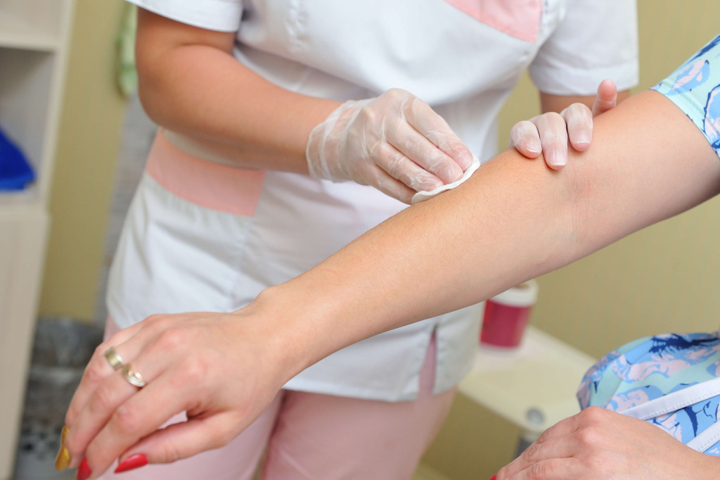
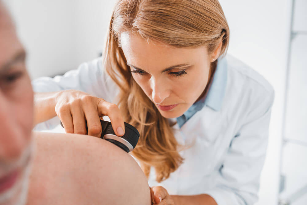
Как подготовиться к процедуре
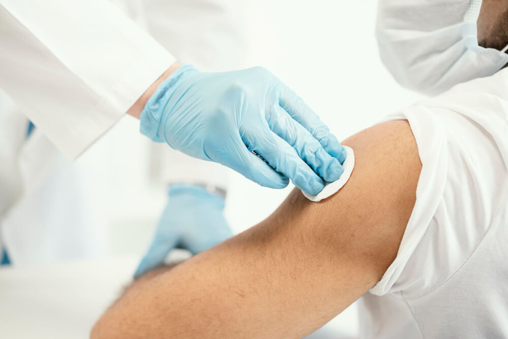
Подготовка к коагуляции очень важна для снижения риска осложнений и достижения максимального эффекта. Обычно она включает несколько этапов:
- Диагностика: проводится осмотр врача-гинеколога или дерматовенеролога с использованием кольпоскопии (для шейки матки и влагалища) или дерматоскопии (для кожи). Дополнительно могут быть назначены анализы: мазок на ВПЧ (для выявления типа вируса), цитология (для исключения дисплазии или рака).
- Консультация: врач определяет подходящий метод коагуляции в зависимости от возраста пациента, локализации кондилом, наличия беременности, сопутствующих заболеваний и иммунного статуса.
- Подготовительные меры: за несколько дней до процедуры рекомендуется избегать солнца и солярия, не использовать агрессивные кремы и мази, а также соблюдать гигиену интимной зоны без спиртовых или раздражающих средств.
Процесс коагуляции
Каждый вариант данной методики имеет свои особенности:
- Химическая коагуляция: наносится аппликатором на пораженные участки, через 5–7 дней ткань отмирает и отторгается. Обычно требуется 2–3 процедуры, между которыми выдерживается интервал в 5–7 дней.
- Лазерная коагуляция кондилом: лазер испаряет патологические ткани бесконтактно и точечно. Процедура занимает 5–15 минут, не требует госпитализации. Одновременно происходит обеззараживание лазерным лучом, что снижает риск инфицирования.
- Радиоволновая коагуляция. Аппарат «Сургитрон» или «Фотек» воздействует на пораженные ткани тепловой энергией, при этом здоровые участки почти не затрагиваются. Благодаря высокой точности рана заживает быстро, а образование рубцов минимально. Ранка обычно покрывается тонкой корочкой, которая отпадает через несколько дней.
- Электрокоагуляция применяется при более плотных и объемных кондиломах. Ток высокой частоты вызывает глубокое прижигание тканей, что позволяет провести гистологический анализ удаленного материала. Минус — риск повреждения здоровой кожи и более выраженный дискомфорт.
Реабилитация
После процедуры коагуляции пациенту важно правильно ухаживать за обработанными участками для скорейшего заживления:
- После лазерной или радиоволновой коагуляции образуется корочка, которая отпадает самостоятельно в течение 7–14 дней. Полное восстановление может занимать от 1 до 4 недель, в зависимости от индивидуальных особенностей организма и выбранного метода.
- Рекомендации: не сдирать корочку, избегать использования агрессивных моющих средств (гелей, мыла), носить хлопковое белье, воздерживаться от половых контактов и горячих ванн минимум 1–2 недели.
- Возможные ощущения: легкая болезненность, покраснение, небольшие кровянистые выделения считаются нормой.
- Когда обращаться к врачу: если появилась сильная боль, температура выше 38 °C, выраженное кровотечение или гнойные выделения — это повод для немедленной консультации.
Коагуляция кондилом — это современный и эффективный метод удаления новообразований, который требует правильной подготовки, выбора подходящей методики и соблюдения рекомендаций врача. Благодаря разнообразию методов коагуляция подходит для большинства пациентов и позволяет добиться оптимальных результатов с минимальными рисками.
Преимущества метода
Коагуляция кондилом — один из самых востребованных способов удаления новообразований, поскольку он сочетает в себе безопасность, эффективность и минимальный риск осложнений. Рассмотрим его ключевые преимущества более подробно.
- Минимальное повреждение рядом расположенных тканей.
- Быстрое восстановление.
- Низкий риск осложнений.
- Отсутствие шрамов и рубцов.
- Возможность гистологического анализа после электрокоагуляции.
- Доступность для разных категорий пациентов.
Особенно заметно это при лазерной и радиоволновой коагуляции. Эти методы воздействуют точно на патологическую ткань, не задевая здоровую слизистую. Благодаря точечному направлению энергии удается избежать ожогов и травмирования, что особенно важно при расположении кондилом на чувствительных участках (влагалище, шейка матки, анальный канал).
Пациенты могут вернуться к обычному образу жизни через 1–2 недели. После радиоволновой коагуляции эпителизация ранки происходит еще быстрее — примерно через 3 дня. Это особенно важно для активных людей, которые хотят минимизировать выпадение из привычного графика. Также коагуляция обычно не требует госпитализации и легко переносится даже амбулаторно.
Современные методы коагуляции (лазер, радиоволны) обеспечивают бесконтактное воздействие, что практически исключает риск инфицирования. Сосуды коагулируются сразу во время процедуры, что предотвращает кровотечение и минимизирует отёчность. Благодаря этому пациенты редко сталкиваются с воспалением или длительным заживлением.
Лазерная и радиоволновая коагуляция способствуют естественному заживлению слизистой без образования грубых рубцов. Это особенно важно для женщин, планирующих беременность или заботящихся о косметическом результате. Отсутствие шрамов также снижает риск психологического дискомфорта.
При использовании электрокоагуляции врач может взять биоптат (образец ткани) для последующего гистологического исследования. Это особенно актуально при подозрении на дисплазию или онкологические процессы, когда важно подтвердить диагноз и выбрать тактику лечения.
Химическая коагуляция остаётся одним из самых бюджетных вариантов. При условии контроля врача препараты могут применяться даже амбулаторно или в домашних условиях (например, Солковагин, Солкодерм). Такой подход особенно удобен для пациентов, которые по разным причинам не могут воспользоваться более дорогостоящими методами.
Таким образом, коагуляция кондилом сочетает в себе ряд преимуществ: безопасность, быстрое восстановление, минимальный риск осложнений и отличный косметический результат. Это делает метод востребованным и удобным для большинства пациентов.
Вопросы и ответы
Можно ли проводить коагуляцию при беременности?
Химические, электрокоагуляционные методы во время беременности не рекомендуются из-за риска ожогов и травм.
Лазер и радиоволны могут применяться во II–III триместре в специализированных клиниках, осторожно и при наличии показаний — на усмотрение врача.
Какие противопоказания существуют?
Выделим основные:
- Острые инфекции, воспалительные процессы в зоне вмешательства;
- Онкологические и предраковые состояния;
- Аллергические реакции на анестезию или химический состав;
- Имплантированные кардиостимуляторы при радиоволновой технике (ограничения условные).
Как долго длится процедура?
Химическая коагуляция занимает несколько минут, эффект развивается через 5–7 дней, всего нужно 1–3 сеанса.
Лазер/радиоволна/электрокоагуляция: от 5 до 40 минут на сеанс, включая подготовку, анестезию и последующую обработку.
Сколько стоит коагуляция?
Цены зависят от метода, места, опыта врача и количества кондилом:
- Химическая коагуляция: в среднем 1 500–3 000 ₽ за сеанс, до 3 образований.
- Лазерная – от 1 500–2 200 ₽ за одну единицу, до 15 000–20 000 ₽ за множественные удаления (6–15 штук).
- Радиоволновая коагуляция: 2 200–5 000 ₽ за штуку; при большом числе — 10–15 000 ₽.
- Электрокоагуляция: 2 000–5 000 ₽, может включать гистологию.
Коагуляция кондилом — современный, эффективный и безопасный способ удаления новообразований, особенно в зоне интимных органов. Лазерная и радиоволновая коагуляция, сочетающие высокую точность, бескровие и быстрый период восстановления, являются приоритетными методами. Они исключают появление рубцов и минимизируют риск рецидива. Химическая коагуляция выгодна по стоимости, но требует строгого профессионального контроля, чтобы предотвратить ожоги и травмы.
Выбор метода зависит от индивидуальных особенностей пациента: локализации кондилом, их размера и состояния слизистой, ожиданий после процедуры, планов беременности. Независимо от метода после удаления следует проводить комплексную профилактику ВПЧ: иммуномодуляция, барьерная контрацепция, вакцинация, регулярный цитологический контроль.
Удаление кондилом — это только часть терапии. Чтобы предотвратить рецидивы и снизить вирусную нагрузку, важны комплексный подход и соблюдение медицинских рекомендаций.


