про услугу
Пайпель-биопсия — это современный способ диагностики состояния эндометрия, который сочетает высокую информативность и минимальную травматичность.
Что такое пайпель-биопсия эндометрия
Пайпель-биопсия — это щадящий и информативный способ исследования слизистой оболочки матки, при котором врач получает небольшой фрагмент эндометрия для анализа. Процедура проводится с помощью тонкой гибкой трубочки (пайпель-катетера), без расширения шейки матки и чаще всего — без обезболивания. Однако при низком болевом пороге может использоваться местная анестезия, особенно у нерожавших женщин.
Проще говоря, это аспирационный метод диагностики, который позволяет «всосать» небольшой участок ткани и изучить его под микроскопом. Такое исследование помогает определить наличие воспаления, атипичных клеток, гиперплазии, полипов и других изменений, влияющих на женское здоровье.
Когда назначают пайпель-биопсию
Процедуру назначают при подозрении на патологии эндометрия — внутренней оболочки матки. Она помогает выявить гормональные нарушения, воспаления, новообразования и предопухолевые изменения. Также её проводят в рамках контроля состояния после хирургических вмешательств и гормональной терапии. Врач может направить на пайпель-биопсию при:
- нерегулярных или болезненных менструациях;
- межменструальных кровотечениях;
- планировании беременности и ЭКО;
- длительном бесплодии без очевидной причины;
- подозрении на гиперплазию, полипы или рак эндометрия;
- хронических воспалениях слизистой матки.
Метод ценится за свою информативность, минимальную травматичность и возможность проведения в амбулаторных условиях без наркоза.
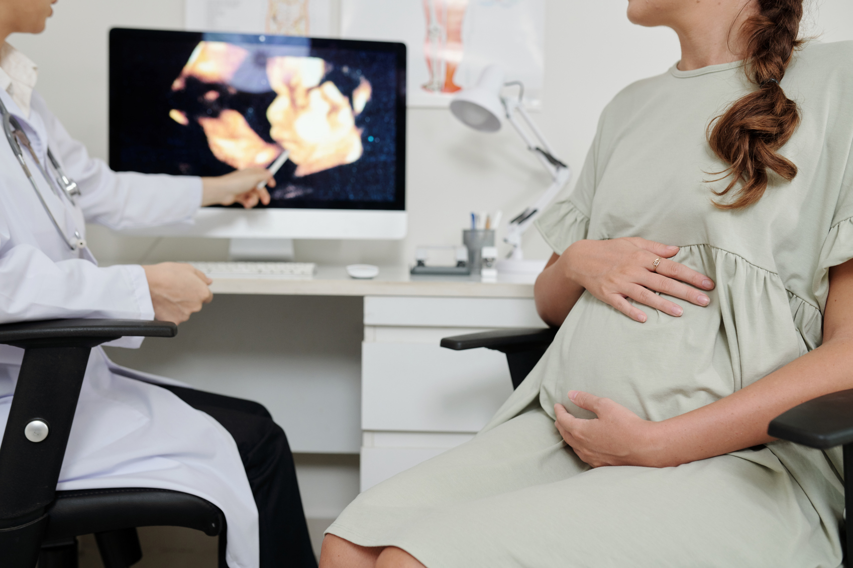
При бесплодии и неэффективном ЭКО
Пайпель-биопсия — важный этап обследования при репродуктивных трудностях. Когда женщина не может забеременеть в течение года или проходит через неудачные попытки ЭКО, врачи ищут скрытые причины. Один из возможных факторов — хронический эндометрит, который часто протекает бессимптомно, но мешает имплантации эмбриона.
Биопсия также позволяет исключить или подтвердить структурные изменения слизистой, гиперплазию или гормональный дисбаланс, которые могут мешать наступлению беременности. По результатам гистологии врач подбирает персонализированную тактику лечения, повышающую шансы на успешное зачатие.
Кровянистые выделения и нерегулярные месячные
Изменения характера менструации — повод для детального обследования. Обильные или слишком скудные, нерегулярные или полностью исчезнувшие месячные, межменструальные мажущие выделения могут быть симптомами нарушений гормонального фона, наличия полипов, гиперплазии или атрофии эндометрия.
Пайпель-биопсия позволяет получить ткань эндометрия для гистологического анализа, выявить наличие воспаления, избыточного разрастания или других патологий.
Гистология выявляет предопухолевые изменения на ранней стадии, когда лечение наиболее эффективно. Метод позволяет избежать ненужного хирургического вмешательства, если изменения доброкачественные, либо вовремя начать терапию при обнаружении злокачественных клеток.
Хронический эндометрит и гиперплазия
Хронический эндометрит развивается после инфекций, выскабливаний, родов или абортов и часто остаётся незамеченным. Он может провоцировать сбои цикла, боли, мажущие выделения и снижать способность к зачатию. Биопсия помогает установить точный диагноз, что особенно важно при планировании беременности.
Гиперплазия — это утолщение слизистой, вызванное гормональным дисбалансом. Она может быть простой, без атипии, либо сопровождаться атипичными клетками — предвестниками рака. Пайпель-биопсия — основной способ дифференцировать эти состояния и вовремя начать лечение.
Однако при подозрении на очаговую атипическую гиперплазию метод может быть недостаточно информативен — тогда прибегают к гистероскопии с прицельной биопсией.
Контроль после операций и лечения
После удаления полипов, проведения выскабливаний или гормональной терапии важно оценить, как восстанавливается эндометрий. Пайпель-метод позволяет это сделать без повторных травм и госпитализации. Он помогает определить эффективность лечения и необходимость его корректировки.
Также биопсия назначается после терапии гиперплазии или эндометрита, чтобы убедиться в отсутствии патологических изменений и следов воспаления. Это важный этап контроля, особенно при подготовке к беременности или перед повторной попыткой ЭКО.
Когда нельзя проводить пайпель-биопсию
Несмотря на безопасность и простоту, пайпель-биопсия имеет ряд противопоказаний. Процедура не проводится при:
- подтвержденной беременности — вмешательство в полость матки может привести к выкидышу или другим осложнениям;
- острых воспалительных заболеваниях органов малого таза — например, при эндометрите, сальпингоофорите, цервиците, так как забор материала может усилить воспаление и привести к его распространению;
- обильных маточных кровотечениях — в таких условиях сложно получить качественный образец, к тому же повышается риск осложнений;
- стенозе (сильном сужении) шейки матки — это затрудняет прохождение катетера и делает процедуру болезненной или невозможной.
- атрезии цервикального канала у пожилых женщин.
- Диагностика: перед биопсией обязательно проводят УЗИ органов малого таза, чтобы уточнить толщину и структуру эндометрия. Также назначаются мазки на флору и инфекции (ИППП, бактериальный вагиноз), общий анализ крови и коагулограмма — для исключения воспалений и нарушений свертываемости.
- Исключение вагинальных вмешательств: за 2–3 дня до процедуры следует избегать половой жизни, не использовать вагинальные свечи, кремы, спринцевания и тампоны. Это снижает риск заноса инфекции и не искажает результаты анализа.
- Коррекция приема лекарств: если вы принимаете антикоагулянты (например, варфарин, аспирин, клопидогрел), обязательно сообщите об этом врачу. Возможно, за несколько дней до биопсии потребуется изменить схему лечения.
- незначительных тянущих болей внизу живота;
- мажущих кровянистых выделений в течение 1–3 дней.
- воздержаться от половой жизни;
- не использовать тампоны, спринцевания, вагинальные свечи;
- избегать тепловых процедур: не посещать баню, сауну, не принимать горячую ванну.
- фазе менструального цикла (пролиферативная, секреторная). Это особенно актуально при диагностике лютеиновой недостаточности и уточнении причин бесплодия;
- наличии воспалительных изменений (острый или хронический эндометрит);
- структуре эндометрия — нормальной или гиперплазированной, с атипичными клетками;
- присутствии полипов или других новообразований;
- предраковых или злокачественных изменениях.
- минимальная травматичность — слизистая практически не повреждается;
- нет необходимости в общем наркозе и госпитализации;
- быстрое выполнение и восстановление;
- высокая информативность, особенно при подозрении на хронический эндометрит, гиперплазию или атипию клеток;
- возможность многократного проведения — например, для контроля эффективности лечения.
- не позволяет точно диагностировать очаговые образования (например, маленькие полипы или участки эндометриоза, которые не попали в образец);
- в редких случаях объем ткани оказывается недостаточным для полноценной оценки;
- возможны болевые ощущения и дискомфорт, особенно у нерожавших женщин или при узком цервикальном канале.
- обильные кровянистые выделения;
- боли внизу живота, не проходящие более суток;
- инфекционные осложнения, особенно при несоблюдении стерильности или наличии скрытых воспалений;
- реакция на антисептики или местное обезболивание (аллергия, раздражение).
Также существует ряд относительных противопоказаний, при которых решение принимается индивидуально. Например, при нарушениях свертываемости крови, после недавних операций или на фоне приема некоторых препаратов. Врач обязательно оценивает общее состояние пациентки, результаты анализов и УЗИ перед тем, как рекомендовать биопсию.
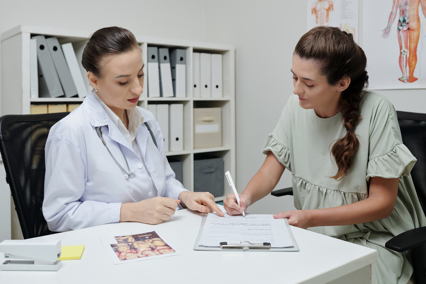
Как подготовиться к процедуре
Чтобы процедура прошла максимально безопасно и информативно, важно правильно подготовиться. Обычно подготовка включает несколько этапов:
В день процедуры не требуется корректировать питание или принимать какие-либо препараты. Однако стоит надеть удобную одежду и взять с собой прокладку — возможны легкие кровянистые выделения после манипуляции.
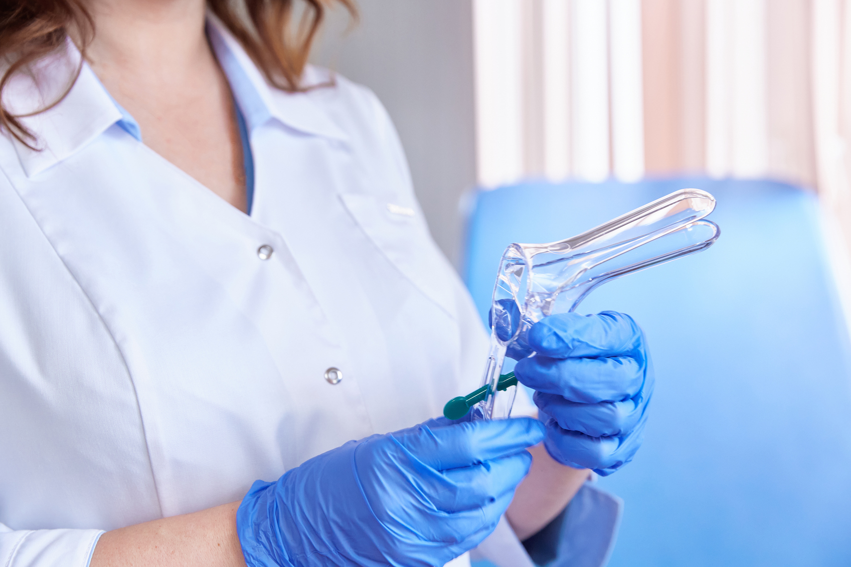
Как проходит пайпель-биопсия эндометрия
Пайпель-биопсия — это амбулаторная процедура, которая проводится без наркоза, чаще всего в кабинете гинеколога. Госпитализация и последующая реабилитация не требуется.
Женщина располагается в гинекологическом кресле. Врач проводит обработку наружных половых органов и шейки матки антисептиком. Затем в шейку матки аккуратно вводится специальный инструмент — пайпель-катетер. Это тонкая гибкая трубка с поршнем внутри.
При введении катетера пациентка может испытывать лёгкий дискомфорт или ощущение кратковременного спазма, похожего на болезненные ощущения при месячных. Обезболивание обычно не требуется, но в случае повышенной чувствительности врач может использовать местный анестетик.
После получения материала катетер извлекается, и пациентка может сразу отправляться домой. Вся процедура, от подготовки до завершения, занимает не более 10–15 минут.
Как берётся материал
Пайпель-катетер устроен по принципу шприца. Когда врач оттягивает поршень назад, создается отрицательное давление (вакуум), за счёт чего внутрь трубочки втягиваются микроскопические фрагменты слизистой оболочки матки — эндометрия.
Это аспирационный метод, то есть ткань не вырезается, а как бы «всасывается» внутрь инструмента. Такой подход снижает риск травмы и инфекции, а также не требует расширения шейки матки, как при классическом выскабливании.
Полученный материал помещается в специальную пробирку с консервирующей жидкостью и направляется в лабораторию на гистологическое исследование. Там под микроскопом патологоанатом оценивает структуру ткани, наличие клеточных изменений, воспалений, полипов и других патологий.
После процедуры: что ожидать
Сразу после манипуляции женщине рекомендуется немного отдохнуть (15–30 минут). Возможно появление:
Эти симптомы считаются нормальными и не требуют лечения.
В течение ближайших 48 часов желательно соблюдать следующие рекомендации:
Если появляются следующие тревожные симптомы — обильное кровотечение, повышение температуры, озноб, гнойные выделения — необходимо незамедлительно обратиться к врачу. Это может свидетельствовать о развитии осложнений.
Что показывает гистология
Результаты гистологического исследования, как правило, готовы в течение 5–10 рабочих дней. Ответ содержит информацию о:
На основе гистологии врач может поставить точный диагноз и назначить соответствующее лечение или дополнительное обследование.
Плюсы и минусы метода
Выделим основные преимущества пайпель-биопсии:
Недостатки и ограничения:
Возможные осложнения
Хотя пайпель-биопсия считается безопасной процедурой, как и любое вмешательство, она имеет риски. В редких случаях могут возникнуть:
Риск осложнений минимален при тщательной подготовке, соблюдении рекомендаций и проведении процедуры опытным специалистом.