про услугу
Что показывает УЗИ желудка
Ультразвуковое исследование желудка - неинвазивный метод диагностики, чаще всего назначаемый детям, а также взрослым пациентам, которые не переносят эндоскопию.
УЗИ позволяет отчетливо увидеть выходной отдел желудка (антральный и пилорический отделы), наиболее близкий к привратнику – месту перехода желудка в двенадцатиперстную кишку, а также начальный ее отрезок.
Иные структуры хуже поддаются визуализации. Однако большая часть поражений желудка возникает именно в выходном отделе, поэтому не стоит недооценивать диагностическую ценность УЗИ.
Показания к УЗИ желудка
Доктор может рекомендовать ультразвуковое исследование желудка пациентам, имеющим противопоказания к ФГДС, а также тем, кто категорически от нее отказывается. Кроме того, УЗИ может применяться в составе комплексной диагностической программы в качестве первичного обследования.
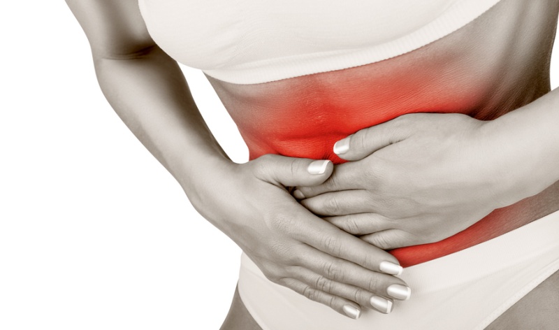
Ультразвуковая диагностика может быть назначена при наличии у пациента следующих симптомов:
-
боли и тяжесть в желудке;
-
снижение аппетита;
-
тошнота и рвота;
-
вздутие живота;
-
частую отрыжка или изжога;
-
сухость или горечь во рту.
УЗИ также может быть информативным при анализе моторно-эвакуаторной функции желудка, оценки функциональности кардиального сфинктера при рефлюксной болезни, а также при подозрении на гастрит, язву, полипы или рак желудка. Еще одно показание - подозрение на стеноз (сужение) антрального и пилорического отделов желудка.
Это лишь часть патологий, которые можно выявить с помощью ультразвука. Лечащий врач также может дополнительно рекомендовать эндоскопию, компьютерную или магнитно-резонансную томографию.
Противопоказания
Ультразвуковое исследование желудка не имеет прямых противопоказаний. Относительными ограничениями могут быть повреждения кожи в области живота, среди которых - ожоги, открытые раны и инфекционные поражения.
Кроме того, применение УЗИ может быть нецелесообразным при избыточном слое висцерального жира (на брюшной стенке).
Как подготовиться к УЗИ желудка
Качественная подготовка - залог максимально информативного исследования.
За 2-3 дня до проведения УЗИ желудка необходимо придерживаться особого рациона, позволяющего снизить газообразование в кишечнике. Так, в списке разрешенных продуктов - нежирная отварная или приготовленная на пару рыба, постное мясо, яйца всмятку, нежирный творог и каши на воде.
При этом лучше исключить темный хлеб, выпечку, кондитерские изделия, сырые овощи и фрукты, черный чай и кофе, алкоголь и газированные напитки, а также бобовые и молочные продукты.
Накануне УЗИ поужинать можно не позднее 19-00-19-30. Тем, кто страдает повышенным газообразованием, стоит пару дней принимать препараты, способные справиться с этой проблемой. Также накануне важно опорожнить кишечник, используя слабительное.
Утром в день процедуры не разрешается не только завтракать, но также употреблять жидкость и курить.
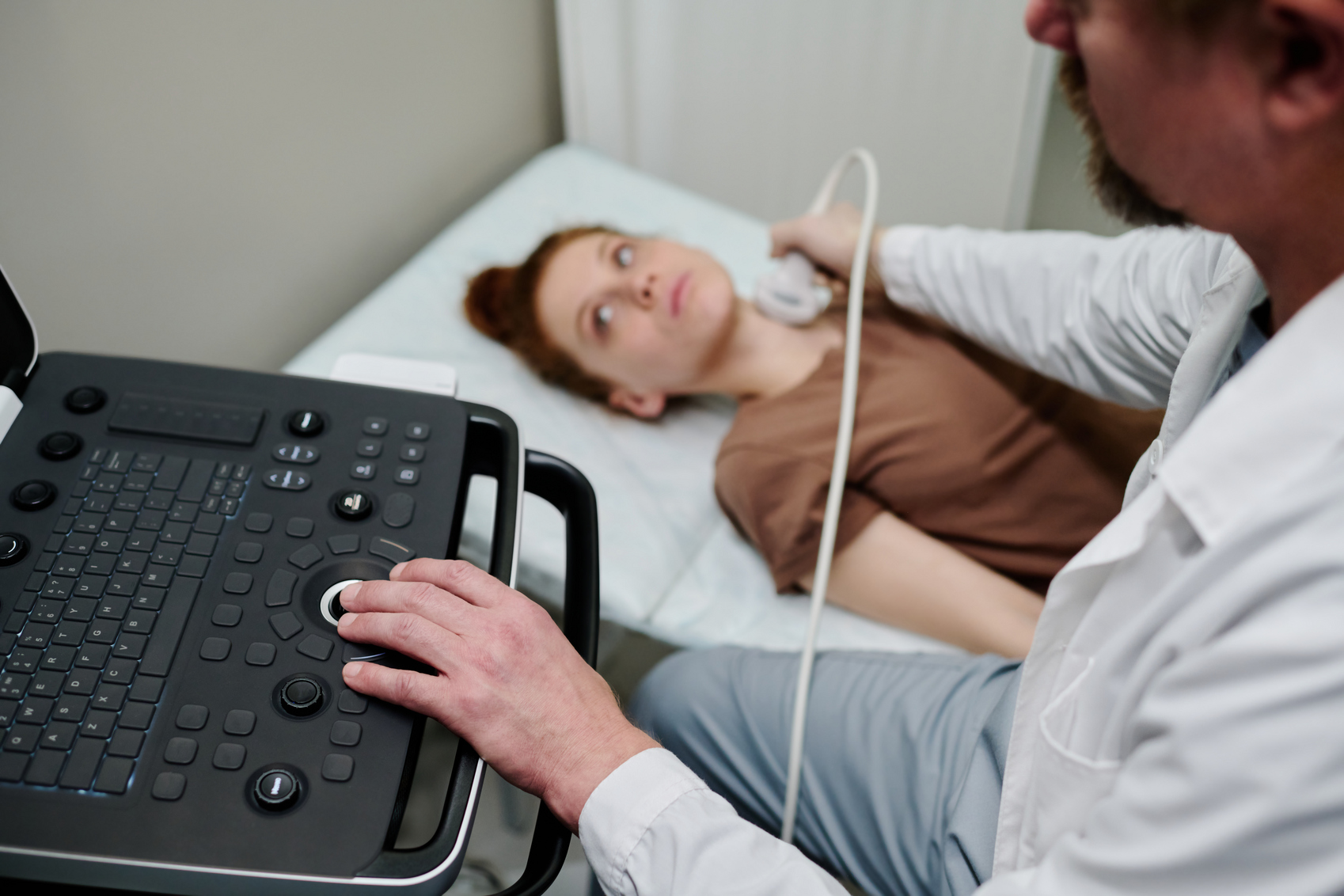
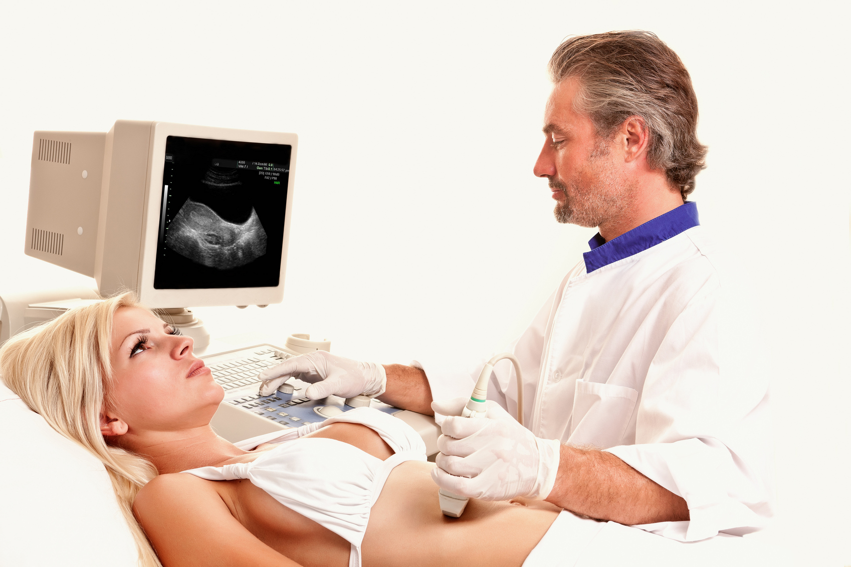
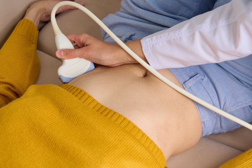
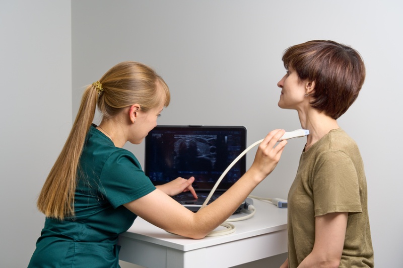
Как проводится УЗИ желудка
УЗИ желудка проводится двумя способами - стандартным через переднюю брюшную стенку (трансабдоминальное УЗИ) и эндо-УЗИ, когда помимо наружного датчика используется миниатюрная камера, размещенная в полости желудка.
Во время исследования пациент лежит на кушетке или находится в полусидячем положении. На его живот наносится гель и специалист начинает перемещать датчик.
Ультразвуковой датчик передает на монитор изображение в нескольких проекциях. Так, доктор оценивает форму желудка и его расположение, а также толщину стенок. Различные деформации и патологические очаги также можно выявить во время УЗИ.
Затем доктор попросит пациента выпить порядка полулитра жидкости, чтобы оценить моторную функцию желудка - то, насколько быстро происходит эвакуация его содержимого.
Эндо-УЗИ – это своего рода комбинация из гастроскопии и традиционного УЗИ. Информацию дает камера, помещенная внутрь, и датчик показывают подробную картину состояния желудка, а также пищевода, двенадцатиперстной кишки, поджелудочной железы и прилегающих органов.
Эндоскопическое ультразвуковое исследование сочетает в себе два метода эндоскопической визуализации с высокочастотным ультразвуком, что позволяет визуализировать стенку желудочно-кишечного тракта и за его пределами, а также близлежащие органы и сосуды. Возможность определить каждый из пяти слоев стенки желудочно-кишечного тракта, соответствующий его гистологическому аналогу, а также обнаружить локализованные лимфатические узлы составляет основу большинства процедур ЭУЗИ.
Показания к эндо-УЗИ можно разделить на несколько категорий:
-
стадирование злокачественных новообразований ЖКТ;
-
оценка панкреатобилиарного заболевания;
-
оценка субэпителиальных аномалий;
-
оценка экстралюминальных аномалий;
-
определение стадии рака легких;
-
терапевтическое ЭУЗИ.
Эндоскопическое УЗИ позволяет сделать биопсию тканей, а также возможно выполнение с его помощью лечебных процедур. В этом его отличие от классической ФГДС.
Стандартное ультразвуковое исследование желудка и кишечника длится 20-30 минут. Затем специалист готовит заключение с предварительным диагнозом.
Преимущества и недостатки УЗИ желудка
Если сравнивать ультразвуковое исследование с классической ФГДС или с КТ, то у первого есть ряд плюсов, а именно:
-
процедура комфортна и безболезненна, поскольку проводится через брюшную стенку;
-
исследование проводится быстро;
-
не создает лучевой нагрузки для пациента, а потому подходит даже детям и беременным женщинам.
Однако УЗИ не лишено и недостатков, среди которых:
-
недостаточно детальное обследование;
-
вероятность пропустить воспалительные и язвенные процессы, а также
-
мелкие опухоли и полипы;
-
невозможность выявить кровоизлияния в стенку желудка;
-
невозможность диагностирования патологических очагов, расположенных в труднодоступных местах.
Кроме того, с помощью УЗИ можно оценить только отдельные сегменты, в то время как другие методы могут давать непрерывное изображение. Также во время ультразвукового исследования нельзя выполнить биопсию подозрительных участков слизистой, определить кислотность желудочного сока, а также выявить наличие или отсутствие Helicobacter Pylori.
Преимущества УЗИ желудка в клинике Expert Clinics
В клинике Expert Clinics в Москве используется высокоточное диагностическое оборудование последнего поколения. А главное, здесь работают доктора, практикующие междисциплинарный подход к коррекции и профилактике различных заболеваний.
Они помогут не только устранить симптомы, но и найдут первопричину болезни, помогут ее устранить, используя персонализированную терапию. В Expert Clinics у вас есть все шансы повысить качество жизни, справившись с заболеваниями желудка.