про услугу
Что такое УЗИ мягких тканей
УЗИ мягких тканей представляет собой неинвазивный метод диагностики и оценки различных состояний и заболеваний мягких тканей тела, таких как мышцы, связки, сухожилия, подкожная жировая клетчатка и кожа.. Этот метод может быть дополнен функцией цветного допплеровского ультразвукового исследования, которая позволяет врачу, проводящему клиническую оценку, также составить исчерпывающий анализ васкуляризации исследуемых органов и обнаружить наличие любых поражений.
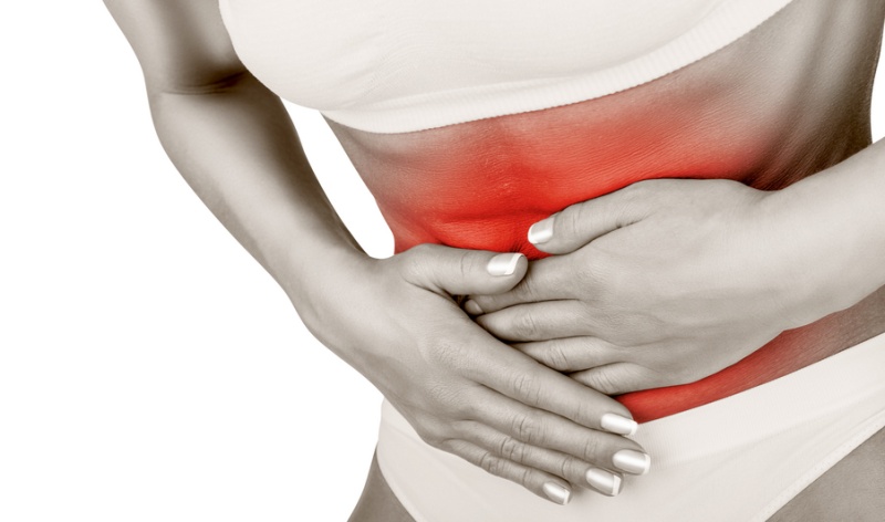
Ультразвуковое исследование обычно назначают с целью анализа доброкачественных лимфатических узлов реактивной или воспалительной природы, а также неопластические. Этот метод медицинского исследования также применяется для выявления и диагностики паховых, брюшных, мошоночных и пупочных грыж.
В большинстве случаев врач назначает УЗИ мягких тканей при обнаружении образования неуточненной природы. Типичным случаем, например, являются поверхностные липомы, доброкачественные образования, которые имеют тенденцию формироваться в жировой ткани и требуют хирургического удаления.
Кроме того, возможно проведение пункционной биопсии под контролем УЗИ. Также с помощью ультразвука доктора наблюдают процесс заживления послеоперационных швов, флегмонов и абсцессов.
Разновидности УЗИ мягких тканей
Ниже приведены некоторые из наиболее распространенных видов ультразвуковой диагностики:
-
УЗИ кожи и подкожной клетчатки
Доктора ежедневно сталкиваются с инфекциями кожи и мягких тканей, а также с инородными телами. Ультразвук дает подробные изображения подкожной и подслизистой ткани и является чрезвычайно полезным инструментом для быстрой оценки этих распространенных проблем. Кроме того, УЗИ информативно для выявления каких-либо аномалий кожи (например, опухолей), а также подкожных отеков и некротизирующего фасциита — он часто связан с предшествующей травмой (например, открытой раной, укусом насекомого), представляет собой редкую, быстро прогрессирующую и опасную для жизни инфекцию, поражающую подкожную клетчатку, фасции и окружающие структуры мягких тканей, включая мышцы.
-
УЗИ суставов
Ультразвуковое исследование назначают при травмах, болезненных ощущениях, отеке и покраснении в области сустава. Ультразвук помогает выявить воспалительные или дегенеративные изменения капсулы сустава, его связок, оценить состояние хрящевых поверхностей, определить наличие выпота (жидкости) в капсуле.
-
УЗИ мышц и сухожилий, фасций и связок
Ультразвуковая визуализация использует звуковые волны для получения изображений мышц, сухожилий, связок и суставов по всему телу. Информативна, например, для диагностики растяжений и переломов.
Позволяет выявлять травмы с повреждением связок, а также воспалительные процессы.
-
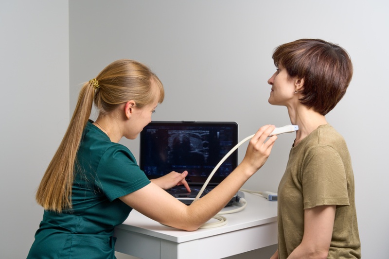
УЗИ мягких тканей головы и шеи
Это ценный метод оценки мягких структур головы и шеи, таких как мышцы, слюнные железы и кровеносные сосуды. Ультразвук может помочь диагностировать такие состояния, как опухоли, инфекции или закупорка кровеносных сосудов.
-
УЗИ паховой области
Это процедура визуализации, нацеленная на область между животом и верхней частью бедра. В первую очередь она используется для осмотра мышц, мягких тканей и выявления распространенных проблем, таких как грыжи (паховые или бедренные), уплотнения или травмы.
-
УЗИ лимфатических узлов
Это метод исследования, который позволяет детально рассмотреть структурные нарушения лимфоузлов, форму и размеры. Диагностика помогает врачу осуществлять контроль лечения длительно текущих и плохо поддающихся лечению хронических заболеваний, например, лимфаденитов или тонзиллитов, а по характерным признакам измененных лимфатических узлов — заподозрить онкопатологию.
УЗИ мягких тканей для диагностики саркомы
Ультразвук очень информативен для диагностики саркомы мягких тканей — формы опухоли, поражающей мышцы, соединительные ткани, жировую ткань, связки, кровеносные и лимфатические сосуды и нервы.
Саркомы мягких тканей могут развиваться как поверхностно, так и глубоко. Часто они протекают бессимптомно, но среди наиболее типичных симптомов можно отметить безболезненный отек.
Для диагностики врачи сначала используют УЗИ мягких тканей, а затем при обнаружении аномальных образований проводят дополнительные исследования.
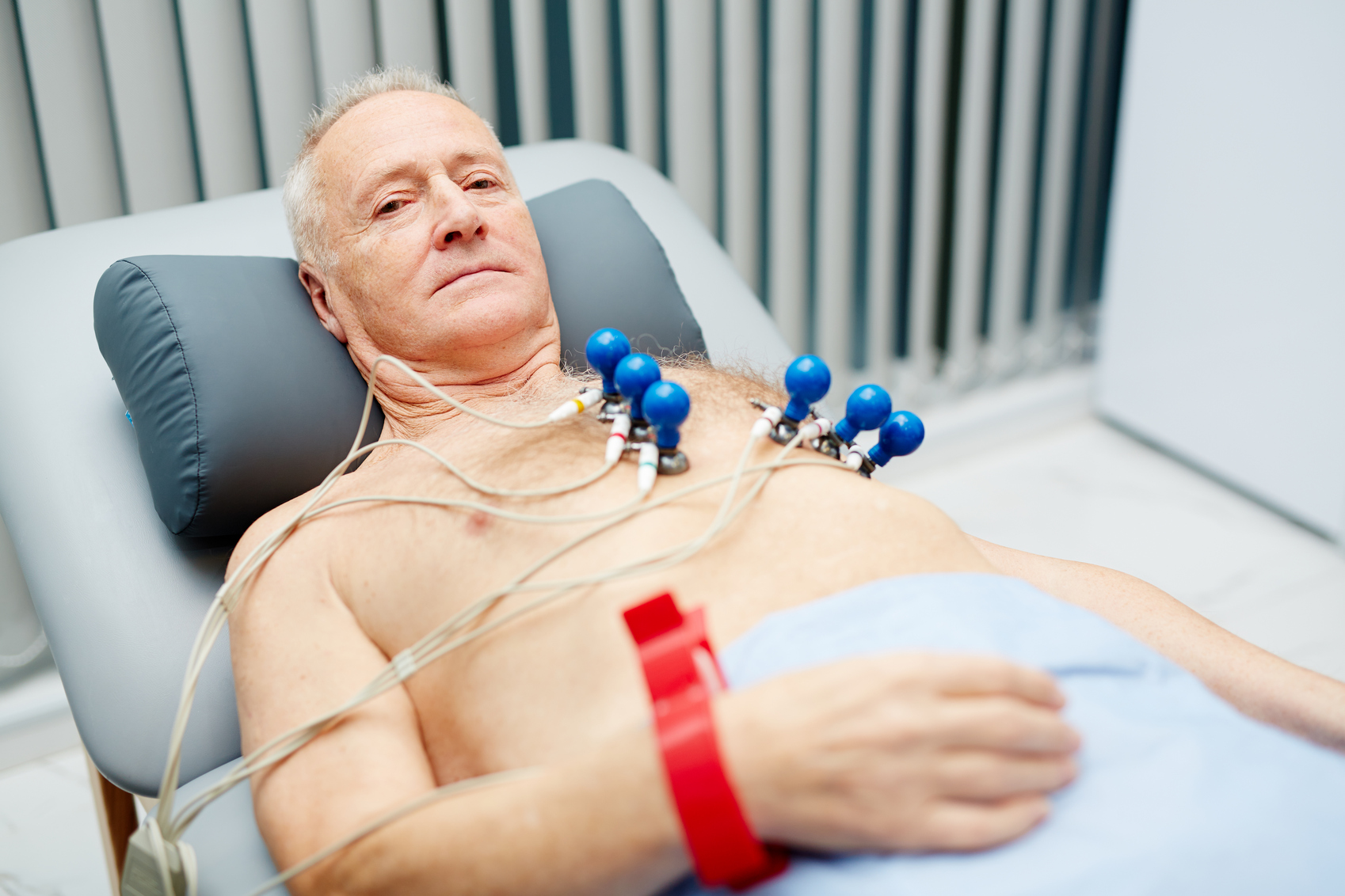
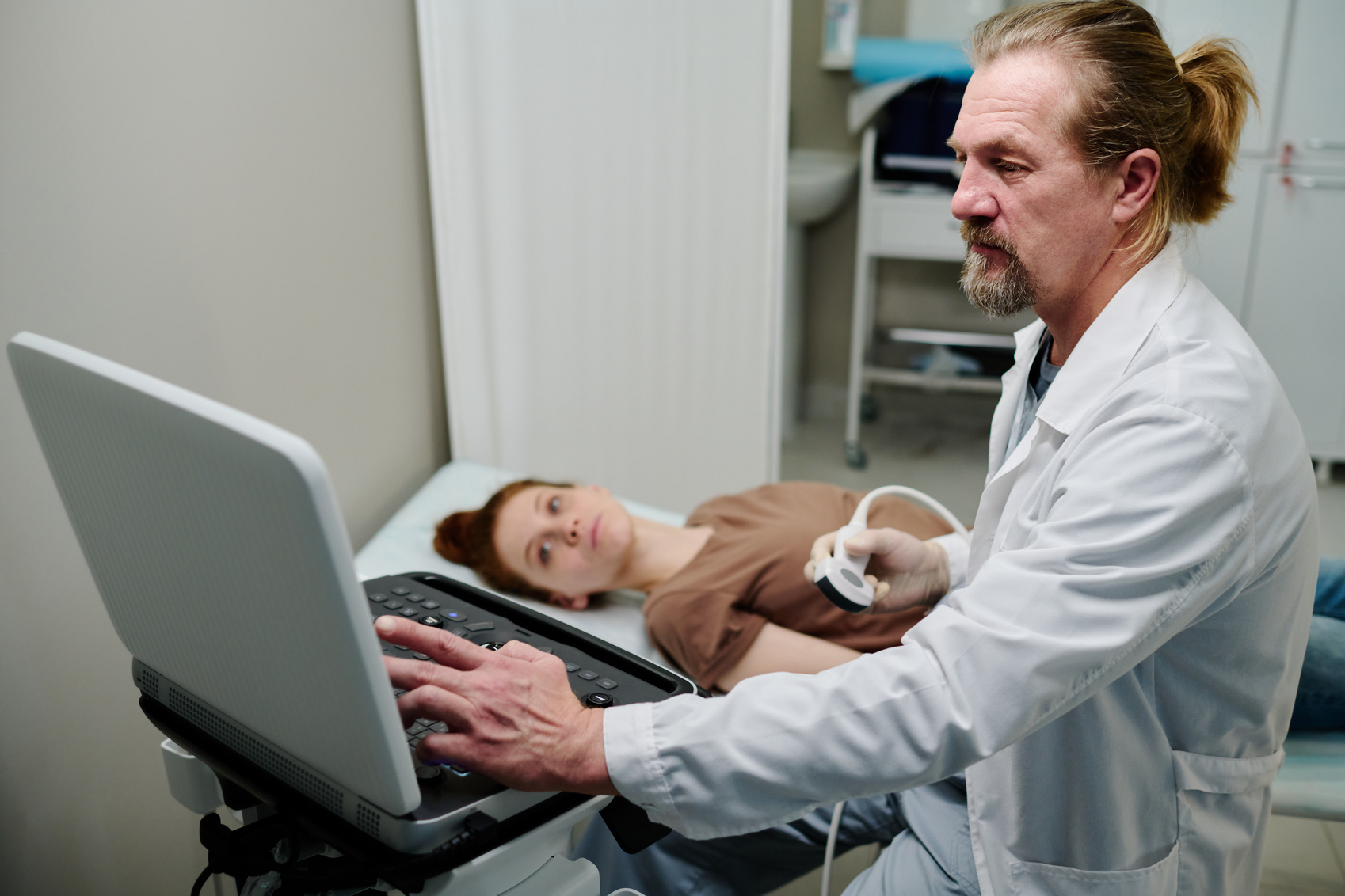
Как проводится УЗИ мягких тканей
Ультразвук мягких тканей — это неинвазивная медицинская процедура, в процессе которой используются высокочастотные звуковые волны для создания изображений структур организма.
Пациент ложится на кушетку и доктор подскажет ему, как расположиться в зависимости от обследуемой области тела. Во время исследования может возникнуть необходимость сменить позицию.
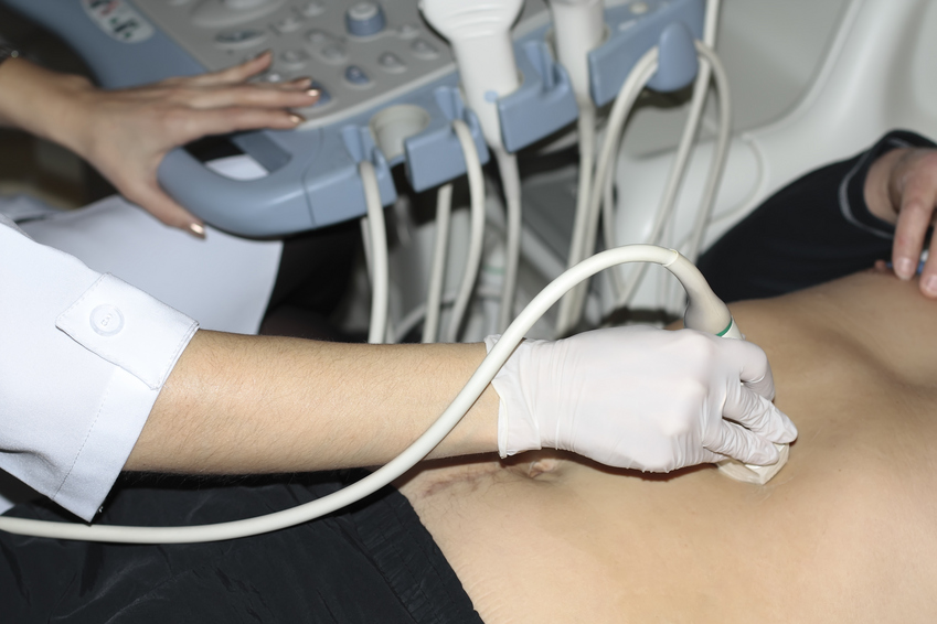
На исследуемую область наносится специальный гель. Он помогает датчику (устройству, которое излучает и принимает звуковые волны) легко скользить и обеспечивает прямой контакт с кожей, устраняя воздушные карманы, которые могут мешать звуковым волнам.
Затем доктор водит датчик по исследуемой области. Это устройство излучает звуковые волны, которые достигают тела и отражаются обратно при столкновении с препятствием (например, органом или тканью). Эти отраженные волны преобразуются в изображения, которые отображаются в реальном времени на мониторе.
Во время процедуры можно сделать фотографии изображений для последующего анализа.
После завершения УЗИ гель удаляется с кожи пациента. Затем специалист выдает заключение.
Как подготовиться
Как правило, для большинства видов УЗИ мягких тканей не требуется специальной подготовки. Однако, в зависимости от области, подлежащей обследованию, пациента могут попросить удалить волосы с соответствующей зоны.
Также рекомендуется приходить на прием к врачу с результатами предыдущих ультразвуковых и других исследований, которые стали причиной обращения. Это поможет доктору составить более ясную и полную диагностическую картину.
Чем заменить УЗИ мягких тканей?
Альтернативных диагностических процедур, доступных и дающих такую же информацию, как УЗИ, не существует. В случае сомнений в характере поражений, обнаруженных при ультразвуковом обследовании, врач может назначить дополнительные инструментальные исследования, такие как компьютерная томография и/или магнитно-резонансная томография.
УЗИ кожи, подкожной клетчатки и мягких тканей представляет собой диагностический метод, который позволяет визуализировать структуры мягких тканей в режиме реального времени и помогает врачам в постановке точного диагноза и выборе оптимальной тактики лечения.
Преимущества УЗИ мягких тканей в клинике Expert Clinics
УЗИ мягких тканей в Expert Clinics проводится врачами с колоссальным опытом в диагностике, в том числе, с точки зрения междисциплинарной медицины.
Также в клинике используется новейшее оборудование, которое позволяет оценить состояние здоровья максимально точно.