про услугу
Анализ соскоба шейки матки — малотравматичное, безболезненное и высокоточное скрининговое исследование. Позволяет выявить онкологическое заболевание на ранней стадии. В каких случаях процедура необходима, а в каких — противопоказана? Как она проводится, что нужно делать до и после взятия соскоба, как долго происходит заживление тканей? Что означают результаты исследования и как их расшифровать? Обо всем этом рассказываем подробно. Объясняем, почему важно проводить диагностику в авторитетных медицинских учреждениях, таких как Expert Clinics.
Как появляются опухоли в шейке матки
Шейка матки состоит из двух частей. Влагалищную, или наружную часть, эктоцервикс, можно исследовать при помощи гинекологического зеркала. Она покрыта многослойным неороговевающим эпителием. Внутренняя часть шейки матки — эндоцервикс, или цервикальный канал — выстлана железистым цилиндрическим эпителием.
Область, в которой ткани эктоцервикса встречаются с клетками эндоцервикса, называется зоной трансформации. Она находится в районе наружного зева. В молодом возрасте этот участок просматривается при помощи гинекологического зеркала, а в пожилом — поднимается вглубь цервикального канала. Именно в этом месте чаще всего появляются злокачественные новообразования. Их появление могут спровоцировать следующие факторы:
-
заражение вирусом папилломы человека (ВПЧ) — онкогенными типами;
-
герпесвирусные инфекции, хламидиоз и другие ИППП;
-
хронические заболевания репродуктивных органов;
-
постоянная смена сексуальных партнеров;
-
раннее начало половой жизни.
Также в группе риска женщины, которые долгое время принимают гормональные средства контрацепции без контроля лечащего врача.
Что такое соскоб шейки матки и для чего он нужен
С момента появления атипичных (измененных) клеток в эпителии половых органов до начала развития злокачественного образования может пройти несколько лет.
Своевременное выявление патологического процесса — залог успешной терапии воспалительных заболеваний органов малого таза.
Гинекологический соскоб (мазок на цитологию, ПАП-тест, мазок по Папаниколау) — информативный и доступный способ диагностики опухолевых патологий. Позволяет вовремя выявить предраковые и раковые заболевания на ранней стадии, определить фоновые воспалительные процессы. Все это позволяет своевременно начать лечение, улучшить прогноз, не допустить развитие новообразования.
Плановая процедура проводится во время профилактического осмотра у гинеколога. Собранные образцы попадают в лабораторию на цитологическое исследование. Специалисты исследуют окрашенный мазок клеток шейки матки на стекле.
Внедрение этого метода диагностики помогло снизить процент заболеваемости раком шейки матки во всем мире.
Показания к проведению диагностики
Забор материала производится во время осмотра у гинеколога. В профилактических целях исследование проводится с определенной периодичностью:
-
каждые три года, если пациентка младше 30 лет;
-
раз в год, если женщина старше 30 лет и находится в репродуктивном возрасте;
-
1–2 раза в год, если присутствуют факторы риска, которые мы перечислили выше (ВПЧ онкогенного типа, ИППП, заболевания репродуктивных органов и др.).
Женщины 65 лет и старше с тремя и более нормальными результатами анализа мазков из шейки матки за последние 10 лет могут не участвовать в скрининге.
Также скрининговое исследование показано при наличии гинекологических проблем, таких как:
-
воспалительные процессы в эндометрии;
-
эрозия (повреждение плоского эпителия) шейки матки;
-
вагинальный дисбактериоз;
-
нарушенный менструальный цикл;
-
бесплодие — невозможность зачатия на протяжении 6 месяцев и дольше;
-
внематочная беременность, невынашивание плода;
-
более двух случаев родов на протяжении 4 лет;
-
кровянистые выделения (не менструации), которые могут появиться после полового акта, особенно после менопаузы;
-
подозрение на ВИЧ, инфекции мочеполовой системы — хламидиоз, микоплазмоз, генитальный герпес и др.;
-
наличие злокачественных образований половых органов у ближайших родственников.
Скрининг проводится при планировании беременности. Обязателен при подготовке к экстракорпоральному оплодотворению. Его проводят в рамках диагностики, для исключения онкологии при гинекологических заболеваний (эрозии шейки матки, воспаления слизистых матки или влагалища, гиперплазии эндометрия и др.) для оценки эффективности лечения.
Противопоказания к проведению скрининга
Процедура имеет ряд ограничений, связанных с состоянием пациентки и наличием некоторых патологий. Получение соскоба с шейки матки противопоказано в следующих случаях:
-
Поздние сроки беременности. Соскоб лучше брать до 22-й недели.
-
Девственность. Девушки, не ведущие половую жизнь, не участвуют в диагностических мероприятиях, так как гинекологические инструменты для скрининга могут повредить девственную плеву.
-
Воспаление половых органов, которое сопровождается сыпью, краснотой, патологическими вагинальными выделениями.
-
Обострение хронических болезней.
-
Терапия урогенитальных инфекций.
-
Вагинальное кровотечение.
Относительными ограничениями являются заболевания, при которых нарушается свертываемость крови.
Подготовка к скринингу
Чтобы исследование дало корректные результаты, к нему нужно правильно подготовиться. Следует исключить факторы, способные исказить результаты.
В рамках подготовки к взятию соскоба рекомендуется:
-
воздержаться от интимной близости за трое суток перед взятием мазка;
-
за неделю до манипуляции не применять вагинальные суппозитории, отказаться от спринцевания;
-
не наносить интимную косметику за 4 дня до диагностики;
-
за 10 суток до соскоба ограничить прием гормональных и антибактериальных препаратов;
-
отказаться от спиртных напитков;
-
накануне провести гигиенические процедуры;
-
непосредственно перед скринингом ограничить употребление жидкости, чтобы не мочиться за 2–3 часа перед взятием мазка.
Также забор материала не производится сразу после кольпоскопии и во время месячных. Лучшее время — не ранее чем на 5-й день менструального цикла и не позднее чем за 5 дней до предполагаемого начала менструации.
Соблюдение этих правил позволяет получить более точные результаты и избежать ошибок в заключении.
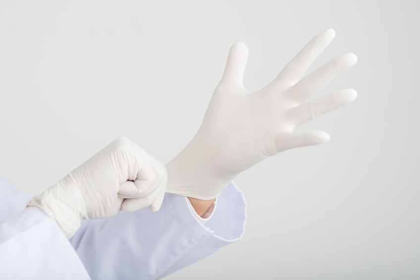
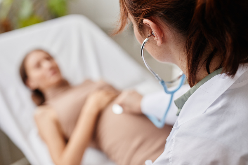
Как проходит взятие соскоба шейки матки
Процедуру проводит врач-гинеколог. Обезболивания не требуется, так как манипуляции не причиняют боли и дискомфорта пациентке.
Соскоб берут из влагалищнной части шейки матки и цервикального канала. Для каждого участка используются отдельные стерильные инструменты. Забор биоматериала с внешней части шейки матки осуществляется при помощи шпателя Эйра (разновидность цервикального шпателя). Для соскоба клеток с поверхности цервикального канала используют жесткую щеточку особой формы — эндобраш.
В тех случаях, когда одновременно проводят биопсию, необходимы электронож и специальные иглы. При этом применяется местная анестезия.
Забор материала для цитологического исследования осуществляется поэтапно:
-
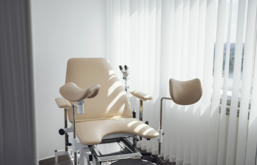
Пациентка размещается в гинекологическом кресле. Выполняется визуальный осмотр половых органов при помощи зеркал. С поверхности эндоцервикса нужно убрать слизь, чтобы открыть доступ к тканям.
-
Далее происходит забор биоматериала с эктоцервикса и эндоцервикса. Гинеколог обнажает шейку матки, аккуратно выскабливает стенку.
-
Образец помещается на предметное стекло.
-
Зону вмешательства (шейку матки) обрабатывают антисептиком. Это необходимо для предупреждения развития инфекции.
Все полученные образцы просушивают на воздухе и отправляют в лабораторию для выявления атипичных клеток. Предварительно врач помечает, с какого участка взят мазок. Применяется следующая маркировка:
-
V — влагалище;
-
C — шейка матки.
Материал наносится на лицевую сторону стекла, маркировка — на оборотную.
Анализ мазка, изучение результатов
Цитологическое исследование соскоба шейки матки осуществляется в условиях лаборатории. Лаборант наносит на стекла зеленку, анилиновые красители, раствор йода. Далее врач-цитолог исследует окрашенный материал и выдает результат в виде заключения.
Сначала специалист оценивает качество полученного биоматериала, после чего изучает клеточный состав. Образец может быть:
-
полноценным — присутствует достаточное количество клеток;
-
недостаточно полноценным — клеточных элементов каждого вида в мазке немного;
-
неполноценным — материала недостаточно для проведения точного анализа.
Существуют несколько методов лабораторного исследования тканей и клеточной структуры эпителия:
-
Микроскопия — изучение клеток, окрашенных особыми веществами, под микроскопом.
-
Использование радиоактивных изотопов. Их помещают в клетки, замещая атомы углерода и кислорода. Это позволяет оценить функционирование клеточных структур.
-
Центрифугирование. Для анализа внутриклеточных структур применяется центрифуга, которая делит биоматериал на отдельные органеллы. Метод актуален для иммуноцитохимического исследования соскобов с шейки матки (жидкостная цитология). Позволяет обнаружить специфические белки и выявить онкопроцесс на начальной стадии.
Интерпретация полученных данных — сложный процесс, требующий знания научных стандартов и высокой квалификации. Поэтому расшифровкой данных и составлением заключения занимаются высококвалифицированные специалисты.
Для расшифровки результатов применяют несколько подходов.
Классы цитологической картины по Папаниколау:
-
1-й. Патологических изменений не имеется.
-
2-й. Присутствуют маркеры, указывающие на наличие инфекционного процесса.
-
3-й. Обнаружена трансформация клеток эпителия. Выделяют три степени — легкая, умеренная, тяжелая дисплазия.
-
4-й. Выявлены отдельные клетки с явными признаками озлокачествления, что свидетельствует о предраковом состоянии, злокачественной опухоли на ранней стадии развития.
-
5-й. Присутствует большое количество злокачественных клеток, что указывает на наличие злокачественного новообразования.
Современные лаборатории используют классификацию Бетесда:
-
NILM (1-й класс) — соответствует норме. Патологических поражений и злокачественных процессов не имеется, лечения не требуется.
-
ASC, ASC-US, ASC-H (2-й класс) — присутствуют реактивные изменения, что может говорить о наличии воспалительного процесса. Его нужно купировать.
-
LSIL (3-й класс) — сигнализирует о наличии дисплазии легкой степени, наличии ВПЧ. Вероятность развития онкологии низкая, но требуется контроль.
-
HSIL (3–4-й класс) — умеренная или тяжелая дисплазия, есть риск трансформации в рак шейки матки. Необходима дополнительная диагностика.
-
CIS (5-й класс) — наличие инвазивного рака. Необходимо срочное лечение.
Если есть даже небольшое подозрение на злокачественное образование, необходимо провести обследование на онкогенные типы ВПЧ.
Реабилитация
После взятия соскоба шейки матки доктор дает рекомендации, что можно и нельзя делать после манипуляции.
Процедура малоинвазивная, травматичность низкая. Тем не менее, в первые 1–2 дня могут появиться кровянистые выделения. Для быстрого восстановления слизистой оболочки следует избегать интимной близости, не пользоваться гигиеническими тампонами. Противопоказаны тепловые воздействия (баня, сауна), купание в водоемах, а также физические нагрузки в первые пару дней после процедуры.
Если процедура проводится в стерильных условиях, опытным специалистом, негативные реакции организма исключены.
Высокоточная диагностика в Expert Clinics
Expert Clinics — клиника европейского уровня в Москве. Мы работаем в области антивозрастной, регенеративной и эстетической медицины. Оказываем широкий спектр медицинских услуг, в том числе осуществляем диагностику и лечение гинекологических заболеваний. В нашей клинике вы можете провести исследование соскоба с шейки матки — сдать мазок и получить расшифровку результата в течение 3-4 рабочих дней.
Высококлассные специалисты нашей клиники будут сопровождать вас на всех этапах диагностики и лечения. Наличие мощной лечебно-диагностической базы, высокая квалификация и опыт наших врачей позволят выявить заболевания на ранней стадии. Это значительно увеличит шансы на успешное лечение.
В Expert Clinics диагностическая процедура выполняется опытными специалистами, с использованием передового оборудования и стерильного, безопасного инструментария экспертного уровня. Это обеспечивает качественное и комфортное для пациентов проведение скрининга, гарантирует получение корректного результата, сводит риск осложнения к нулю.
Преимущества обслуживания в нашей клинике:
-
Высокое качество проведения исследований, быстрота получения результатов, высокая точность.
-
Профессиональная расшифровка данных, подробная консультация с детальным пояснением. При необходимости врач направляет пациентку на дополнительное обследование, на консультацию к профильным специалистам.
-
Протоколы проведения всех лечебных и диагностических процедур соответствуют высоким международным стандартам. Результаты принимаются в отечественных и зарубежных клиниках.
-
Многопрофильная команда опытных докторов с мировым именем, многолетним опытом диагностики и терапии гинекологических заболеваний. Наши врачи постоянно повышают свой уровень на конференциях, симпозиумах в России и за рубежом.
-
Современное диагностическое и лечебное оборудование последнего поколения, соответствующие всем требованиям.
-
Передовые малоинвазивные органосохраняющие методики лечения с доказанной безопасностью и эффективностью.
-
Уютная атмосфера, доброжелательное отношение персонала. Нам дорог каждый пациент, мы окружаем его заботой и вниманием.
Записаться на прием к гинекологу с целью проведения соскоба шейки матки можно прямо на сайте или позвонив по номеру +7 (495) 165-55-95. Уточнить стоимость услуги вы можете у наших менеджеров.
Вопрос/ответ
1) Сколько заживает шейка после цитологии?
В процессе забора материала производится соскоб с помощью щеточки. При этом происходит минимальная травматизация тканей. Они заживают в течение одного-двух дней. В это время можно заметить бледно-розовые выделения из влагалища.
2) Что значит плохой мазок на цитологию?
Если в материале, взятом при соскобе шейки матки, обнаружены атипичные, измененные клетки с признаками озлокачествления, это может свидетельствовать о предраковом состоянии или наличии онкозаболевания. Необходимо проведение дополнительных исследований для уточнения этиологии. При подтверждении диагноза необходимо лечение по показаниям.
Лечение проводится в следующих случаях:
-
Наличие патологически измененных клеток (умеренный дискариоз).
-
Присутствие аномальных клеток, вероятность предрака с высокой степенью злокачественности (тяжелый дискариоз).
-
Присутствие в анализе аномальных клеток, указывающих на наличие инвазивной карциномы.
-
Наличие аномальных железистых клеток, происходящих из слизистой оболочки, шейки матки или яичников (железистая неоплазия).
Исследование может дать отрицательный результат. Это значит, что клетки эпителия соответствуют норме, атипичных клеток, указывающие на дисплазию и другие патологические изменения, не обнаружено. Если цитологическое заключение гласит, что анализы в пределах нормы, лечения не требуется.
Присутствие плоских клеток эпителиального слоя в биоматериале нормально. Если их нет, это может означать атрофию эпителия, дисбаланс мужских и женских половых гормонов. Повышенный уровень лейкоцитов свидетельствует о развитии воспалительного процесса.
3) Что нельзя делать после цитологии шейки матки?
Взятие соскоба — малотравматичная процедура, повреждение тканей незначительное. Чтобы заживление прошло быстрее, в первые дни следует отказаться от половых контактов, спринцевания, использования гигиенических тампонов (следует заменить на прокладки). Также не рекомендуется посещать сауну, баню, плавать в открытых водоемах. Тяжелых физических нагрузок нужно избегать.
СВЯЗАННЫЕ КОМПЛЕКСНЫЕ ПРОГРАММЫ
ВАС МОГУТ ЗАИНТЕРЕСОВАТЬ УСЛУГИ
Воспалительные заболевания женских половых органов занимают первое место во всей структуре гинекологических заболеваний, составляя от 50 до 70% от общего количества случаев.
Подробнее