про услугу
УЗИ органов малого таза благодаря своей безопасности и информативности является одним из главных методов выявления гинекологических и урологических патологий.
Показания к проведению УЗИ
Показания к УЗИ органов малого таза у женщин разнообразны:
- боли в животе различного характера;
- нарушения менструального цикла;
- сильные менструальные боли;
- кровь в кале, моче, вагинальных выделениях;
- нарушения мочеиспускания (слишком частые / болезненные позывы);
- воспалительные процессы;
- подозрение на опухолевые заболевания;
- неприятные ощущения во время интимной близости;
- планирование беременности.
У мужчин к этому списку добавляются еще 4 пункта:
- Профилактика. Она становится обязательной после 40 лет.
- Подозрения на воспаления предстательной железы;
- Боль в области мочевого пузыря;
- Боль в мошонке.
Какие заболевания диагностируются при УЗИ органов малого таза
УЗИ органов малого таза может выявить следующие заболевания:
- очаговые и диффузные изменения во всех слоях матки: полипы эндометрия, наличие гиперплазии, эндометриоз, миома;
- кисты;
- простатит, аденома;
- мочекаменная болезнь;
- доброкачественные и злокачественные опухоли;
- заболевания сосудов яичек, семенников;
- патология кровообращения в органах.
Какие органы исследуют с помощью УЗИ органов малого таза
Это исследование позволяет оценить состояние:
- матки и ее шейки;
- маточного кровотока;
- яичников;
- маточных труб;
- фолликулярного аппарата;
- толщину эндометрия;
- лимфоузлов;
- мочевого пузыря.
У мужчин к мочевому пузырю добавляется предстательная железа и семенные пузырьки.
УЗИ измеряет размеры, расположение, смещаемость органов и их составляющих элементов.
Как проходит исследование
Оно может быть проведено двумя способами. Всё зависит от цели процедуры и жалоб пациента, а также от его индивидуальных особенностей.
-
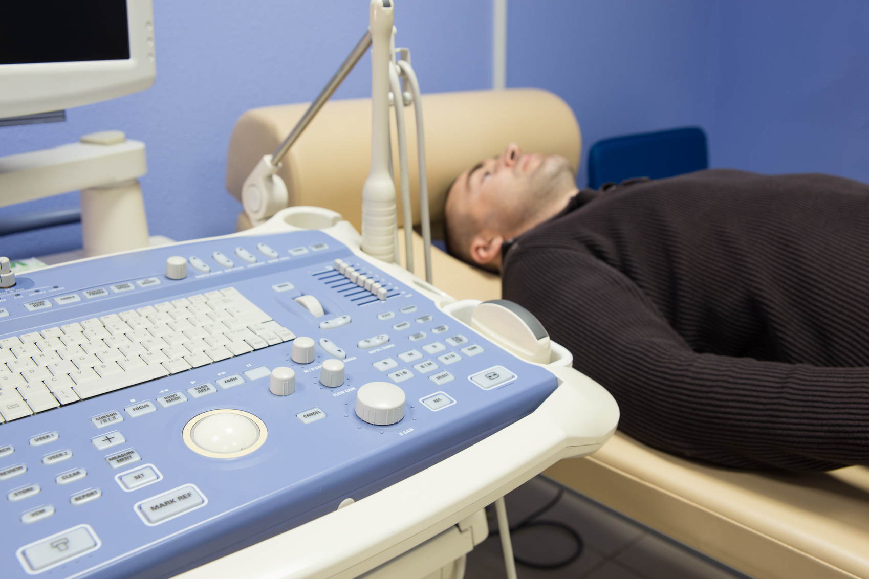
Трансабдоминальное – это классическое УЗИ через переднюю стенку живота, когда пациент ложится на кушетку, а врач с помощью датчика, смазанного специальным гелем, проводит диагностику области снаружи.
-
Трансвагинальное УЗИ хорошо известно женщинам, живущим половой жизнью, так как часто применяется в гинекологии. При наличии девственной плевы такой метод не используется, чтобы не повредить её. Трансвагинальное УЗИ дает более точный анализ органов малого таза. Оптимальное время исследования – это 5-8 день цикла.
Этапы проведения:
-
Пациент снимает бельё, ложится на кушетку.
-
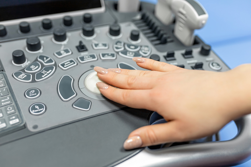
Врач с помощью специального датчика изучает необходимую область.
Подготовка к УЗИ
Прежде, чем сделать УЗИ органов малого таза, стоит обратить внимание на несколько рекомендаций:
- Если вам предстоит трансабдоминальное УЗИ, то необходимо наполнить мочевой пузырь. Для этого следует выпить 1,5-2 литра воды за два часа до исследования и не ходить в туалет.
- Перед трансвагинальным УЗИ необходимо опорожнить мочевой пузырь.
- Независимо от вида исследования, пациенту лучше за 1-2 дня исключить пищу, способствующую газообразованию. К ней относятся картофель, белокочанная капуста, кондитерские изделия, сыр, овощи, фрукты, алкоголь. Повышенное образование газов снижает видимость органов на экране.
Результаты УЗИ
По окончании УЗИ пациент получает развернутую расшифровку результатов. Если в процессе исследования обнаружены патологические нарушения, то врач указывает их структуру, локализацию, размер и другие особенности.
Чтобы точно узнать причину конкретных изменений, врач направит вас к нужному специалисту.
Преимущества УЗИ органов малого таза в Expert Clinics
УЗИ органов малого таза позволяет оценить, насколько хорошо работает репродуктивная система человека. Поэтому женщинам от 20-25 лет и мужчинам от 40 лет крайне важно каждый год проходить это обследование. Оно длится около 15 минут. За это время вы получите ценнейшую информацию о своем здоровье.
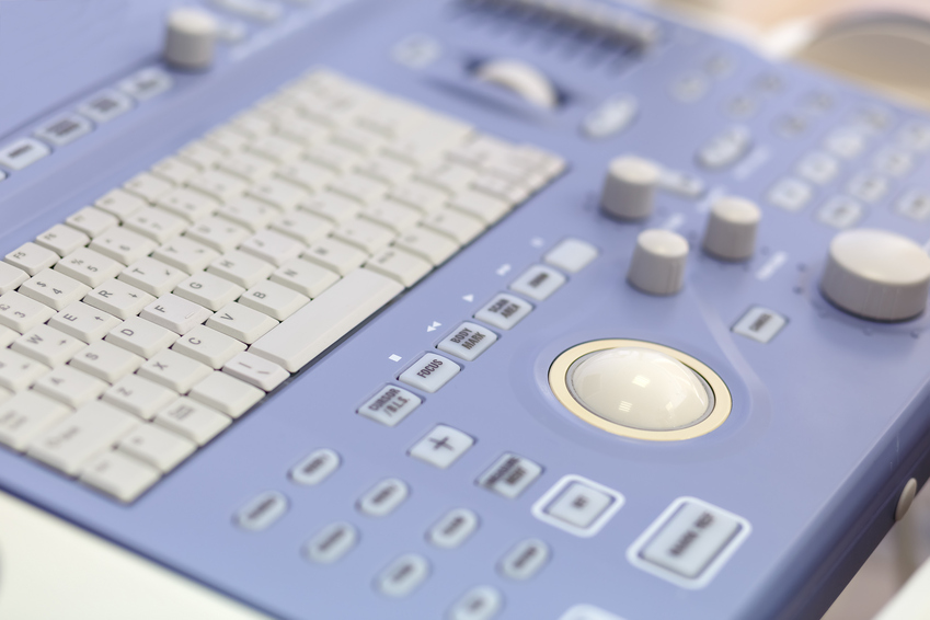
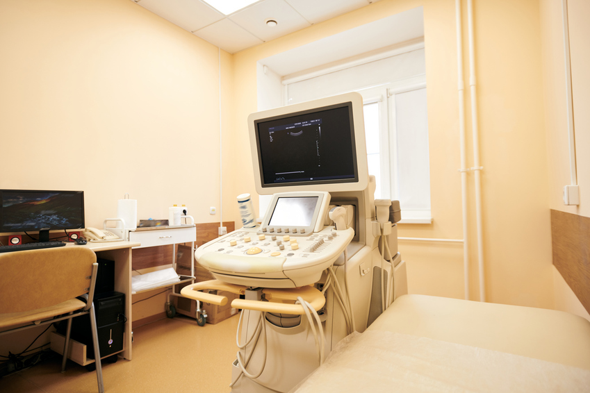
Наша клиника оснащена диагностическим оборудованием последнего поколения. Оно помогает получить высококачественную ультразвуковую картину. Это дает возможность в разных проекциях оценить состояние матки, придатков и других органов.
Врачи Expert Clinics ежегодно проходят повышения квалификации как в России, так и за рубежом, что позволяет нам всегда идти в ногу с современной медициной и применять новые подходы к лечению заболеваний.
К дополнительным плюсам клинки относятся:
-
удобное расположение;
-
запись в подходящее для вас время;
-
индивидуальный подход к каждому пациенту;
-
безопасность, надежность, эффективность диагностики и лечения.