про услугу
Что такое УЗИ кишечника?
Ультразвуковое исследование кишечника - диагностическая процедура, используемая для анализа состояния кишечника, особенно конечного отдела тонкой кишки, аппендикулярной области и толстой кишки.
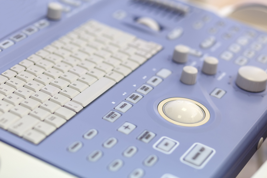
Это неинвазивный метод диагностики, использующий для визуализации ультразвуковые волны. Применяются специальные ультразвуковые датчики с частотой 7,5 МГц и выше в зависимости от телосложения пациента и визуализируемого отдела кишечника.
Наилучшие результаты УЗИ кишечника достигаются при оценке воспалительных изменений в кишечнике при язвенном колите, болезни Крона, при подозрении на аппендицит.
Следует также подчеркнуть, что помимо оценки толщины стенки кишечника и, следовательно, наличия воспалительных или других патологий (например, дивертикулов или новообразований), УЗИ кишечника также позволяет оценить структуры, окружающие кишечник, и выявить наличие лимфатических узлов, возможные свищи и абсцессы.
Что касается аппендикулярной патологии, то в подавляющем большинстве случаев УЗИ позволяет визуализировать воспаленный отросток и любые периаппендикулярные абсцессы.
При дивертикулите УЗИ, помимо выделения места и распространенности воспаления, позволяет контролировать ход медикаментозной терапии и возможное появление осложнений.
Благодаря УЗИ можно оценить не только толщину стенок петель кишечника и, следовательно, состояние воспаления, но и моторику, т. е. движение, и растяжение петель, а также наличие жидкости, что является важным признаком некоторых патологий. В этом случае дополнительно к ультразвуку может быть назначен рентген.
Виды УЗИ
Оценка состояния кишечника посредством ультразвука может проводиться двумя методами.
-
Трансабдоминальное УЗИ. В этом случае исследование проводится через кожный покров живота. И хотя этот способ обычно не доставляет дискомфорта, он недостаточно информативен.
-
Эндоректальное УЗИ. Оно проводится с использованием очень тонкого датчика, который вводится в анальное отверстие. Перед этим прямую кишку наполняют жидкостью, чтобы повысить информативность диагностики и рассмотреть каждую петлю кишечника детально. Манипуляция безболезненна.
-
Трансвагинальное УЗИ. Оно проводится путем введения полостного датчика во влагалище. Этот метод применяется у женщин при подозрении на ректовагинальный свищ для определения свищевого хода и его протяженности. Он также позволяет лучше рассмотреть сигмовидную и прямую кишку при недостаточной подготовке к исследованию.
Лечащий врач может рекомендовать применение нескольких вариантов УЗИ кишечника.
Показания для проведения УЗИ кишечника
УЗИ кишечника проводится для проверки наличия аномалий в петлях кишечника, аппендикулярном отростке и толстой кишке, при подозрении на тромбоз мезентериальных сосудов или ишемию части кишки, кишечное кровотечение, перфорацию кишечной стенки, на наличие свищевого хода или абсцесса и может быть полезно при подозрении на острые и хронические воспалительные заболевания кишечника, такие как:
-
болезнь Крона;
-
дивертикулез или дивертикулит;
-
аппендицит;
-
сегментарный колит, связанный с дивертикулами;
-
неспецифический колит или энтерит.
Ультразвуковое исследование может быть полезно при диагностике неоплазий тонкой кишки, которые относятся к редким опухолям и не имеют характерных симптомов. При проведении УЗИ кишечника возможное обнаружение опухолевых новообразований толстого кишечника. В таких ситуациях УЗИ кишечника является дополнительным методом диагностики и не заменяет колоноскопию или такие исследования, как КТ или МРТ кишечника.
Однозначными показаниями для проведения УЗИ являются также следующие симптомы:
-
неприятный привкус, зловонное дыхание;
-
длительный запор или понос;
-
тяжесть в животе после употребления пищи;
-
изжога, вздутие живота или метеоризм;
-
беспричинная тошнота или рвота;
-
кровь или белая слизь в кале;
-
неприятные ощущения при попытке опорожнить кишечник.
ВАЖНО: Если перечисленные симптомы сопровождаются повышением температуры тела, необходимо обратиться к врачу как можно скорее.
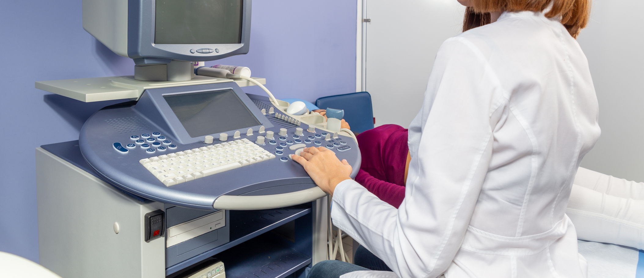
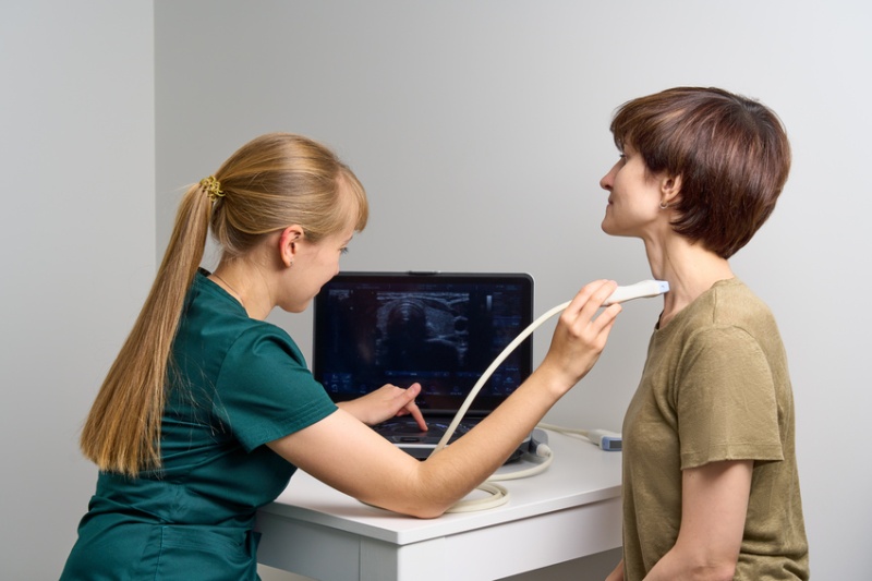
Как проходит УЗИ кишечника
УЗИ кишечника похоже на классическое ультразвуковое исследование брюшной полости: врач наносит гель на живот пациента и благодаря ультразвуковому датчику проверяет наличие утолщений или изменений петель кишечника.
Это безболезненный метод диагностики, за исключением случаев, когда его проводят на воспаленных участках, в этом случае пациент может ощущать небольшой дискомфорт.
Если исследование проводится для оценки петель тонкой кишки, может быть полезно дать больному выпить около литра воды; в то время как, если показанием к тесту является оценка кишечного транзита и перистальтики, исследование также можно проводить с интервалами. Его общая продолжительность в этом случае составит примерно 2 часа.
Стандартное УЗИ кишечника длится примерно 20 минут.
Подготовка к УЗИ кишечника
Данный вид УЗИ проводят натощак, спустя 8-10 часов после последнего приема пищи, и он требует следующей подготовки:
-
Соблюдайте облегченный рацион в течение 3 дней, избегая жареного, жирного, острого;
-
На этот же период откажитесь от употребления алкоголя;
-
Воздержитесь от продуктов, способствующих повышенному газообразованию (бобовые, темный хлеб, цельное молоко, большое количество овощей и фруктов);
-
При склонности к метеоризму воспользуйтесь ветрогонными препаратами;
-
Накануне исследования необходимо сделать очистительную клизму до чистых вод, либо использовать препараты для подготовки к колоноскопии.
Противопоказания
Что касается трансабдоминального УЗИ, прямых противопоказаний для его проведения нет. Его проведение может быть отложено при наличии сильных повреждений кожного покрова (ожоги и травмы).
Ректальное ультразвуковое исследование не стоит проводить при наличии психических расстройств, инфекционных заболеваний кишечника и стеноза терминального отдела желудочно-кишечного тракта.
Преимущества обращения в клинику Expert Clinics
В клинике Expert Clinics в Москве используется высокоточное диагностическое оборудование последнего поколения. А главное, здесь работают доктора, практикующие междисциплинарный подход к коррекции и профилактике различных заболеваний.
Они помогут не только устранить симптомы, но и найдут первопричину болезни, помогут ее устранить, используя персонализированную терапию. В Expert Clinics у вас есть все шансы повысить качество жизни, справившись с болезнями.